Ciftel Sedat, Ciftel Serpil, Altuner Durdu, Huseynova Gulbaniz, Yucel Nurinisa, Mendil Ali Sefa, Sarigul Cengiz, Suleyman Halis, Bulut Seval
Division of Gastroenterology, Erzurum City Hospital, Erzurum, Turkey.
Department of Endocrinology, Faculty of Medicine, Health Science University, Erzurum, Turkey.
BMC Pharmacol Toxicol. 2025 Mar 24;26(1):67. doi: 10.1186/s40360-025-00901-7.
Hepatotoxicity of pyrazinamide, an antituberculosis drug, limits its therapeutic use and oxidative stress has been implicated in this toxicity. This study investigated the protective effects of adenosine triphosphate (ATP), thiamine pyrophosphate (TPP), melatonin, and Liv-52, which have previously been shown antioxidant activities, on pyrazinamide-induced hepatotoxicity.
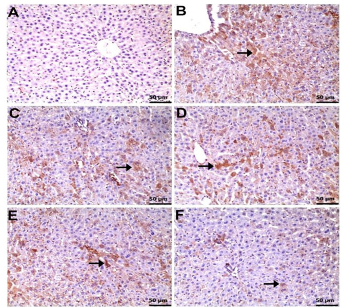
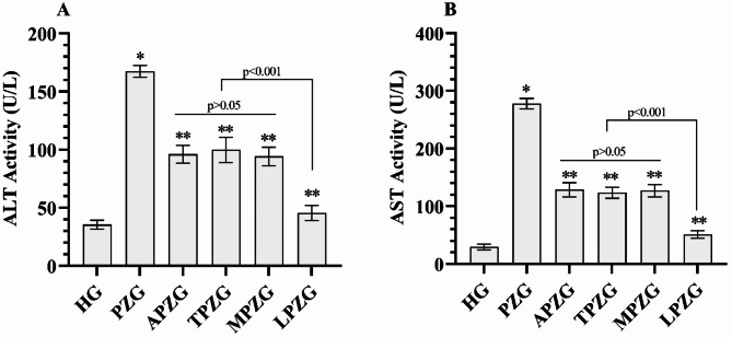
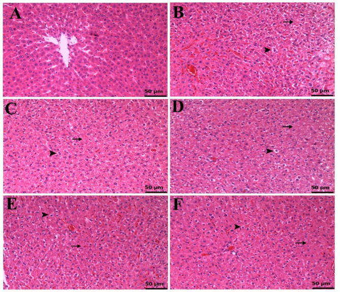
36 albino Wistar male rats were divided into randomized six groups; healthy (HG), pyrazinamide (PZG), ATP + pyrazinamide (APZG), TPP + pyrazinamide (TPZG), melatonin + pyrazinamide (MPZG) and Liv-52 + pyrazinamide (LPZG) groups. ATP 4 mg/kg and TPP 25 mg/kg were administered intraperitoneally (IP). Melatonin 10 mg/kg and Liv-52 20 mg/kg were given orally. One hour after administration of ATP, TPP, melatonin, and Liv-52, 250 mg/kg pyrazinamide was applied orally to all rats except HG group. The treatment was repeated (1 × 1) for 4 weeks. Then, blood samples were taken for determination of alanine aminotransferase (ALT) and aspartate aminotransferase (AST) activities. Immediately after, the rats were euthanized with thiopental sodium (50 mg/kg, IP), and the livers were removed. The tissues were analyzed for malondialdehyde (MDA), total glutathione (tGSH), superoxide dismutase (SOD), and catalase (CAT) also hydropic degeneration, necrosis, and apoptosis (caspase 3) were examined.One-Way ANOVA was used in biochemical analyses and Tukey test was used as post-hoc. For histopathological and immunohistochemical analysis, the Kruskal-Wallis test was used and Dunn's test as a post-hoc.
Pyrazinamide increased MDA land decreased tGSH, SOD, and CAT levels in liver tissues (p < 0.001). It also increased serum ALT and AST activities and caused severe hydropic degeneration and necrosis in liver tissue (p < 0.001). ATP, TPP, melatonin, and Liv-52 significantly prevented the biochemical and histopathological changes induced by pyrazinamide (p < 0.05). On the other hand, Liv-52 was more successful than other potential protectors in protecting liver tissue from pyrazinamide damage (p < 0.05).
ATP, TPP, melatonin, and Liv-52 can be used to protect liver tissue from pyrazinamide-induced hepatotoxicity in rats.
抗结核药物吡嗪酰胺的肝毒性限制了其治疗应用,氧化应激被认为与这种毒性有关。本研究调查了三磷酸腺苷(ATP)、硫胺素焦磷酸(TPP)、褪黑素和利维52(Liv-52)对吡嗪酰胺诱导的肝毒性的保护作用,这些物质先前已显示出抗氧化活性。
将36只白化Wistar雄性大鼠随机分为六组:健康组(HG)、吡嗪酰胺组(PZG)、ATP+吡嗪酰胺组(APZG)、TPP+吡嗪酰胺组(TPZG)、褪黑素+吡嗪酰胺组(MPZG)和利维52+吡嗪酰胺组(LPZG)。ATP 4mg/kg和TPP 25mg/kg腹腔注射(IP)。褪黑素10mg/kg和利维52 20mg/kg口服。在给予ATP、TPP、褪黑素和利维52 1小时后,除HG组外,所有大鼠口服250mg/kg吡嗪酰胺。治疗重复(1×1)4周。然后,采集血样测定丙氨酸氨基转移酶(ALT)和天冬氨酸氨基转移酶(AST)活性。之后,立即用硫喷妥钠(50mg/kg,IP)对大鼠实施安乐死,并取出肝脏。分析组织中的丙二醛(MDA)、总谷胱甘肽(tGSH)、超氧化物歧化酶(SOD)和过氧化氢酶(CAT),同时检查水样变性、坏死和凋亡(半胱天冬酶3)情况。生化分析采用单因素方差分析,事后检验采用Tukey检验。对于组织病理学和免疫组织化学分析,采用Kruskal-Wallis检验,事后检验采用Dunn检验。
吡嗪酰胺增加了肝脏组织中的MDA水平,降低了tGSH、SOD和CAT水平(p<0.001)。它还增加了血清ALT和AST活性,并导致肝脏组织严重的水样变性和坏死(p<0.001)。ATP、TPP、褪黑素和利维52显著预防了吡嗪酰胺诱导的生化和组织病理学变化(p<0.05)。另一方面,在保护肝脏组织免受吡嗪酰胺损伤方面,利维52比其他潜在保护剂更有效(p<0.05)。
ATP、TPP、褪黑素和利维52可用于保护大鼠肝脏组织免受吡嗪酰胺诱导的肝毒性。