Chitsaz Daryan, Rowley Christopher D, Uccelli Nonthué A, Lefebvre Sarah, Krahn Andrea I, Reintsch Wolfgang E, Durcan Thomas M, Tardif Christine L, Kennedy Timothy E
Integrated Program in Neuroscience, McGill University, Montréal, Quebec, Canada.
Department of Neurology & Neurosurgery, Montreal Neurological Institute-Hospital, McGill University, Montréal, Quebec, Canada.
Brain Behav. 2025 Apr;15(4):e70308. doi: 10.1002/brb3.70308.
The common marmoset is a small nonhuman primate that has emerged as a valuable animal model in neuroscience research. Accurate analysis of brain tissue is crucial to understand marmoset neurophysiology and to model neurodegenerative diseases. Many studies to date have complemented magnetic resonance imaging (MRI) with histochemical staining rather than immunofluorescent labeling, which can generate more informative and higher resolution images. There is a need for high-throughput immunolabeling and imaging methodologies to generate resources for the burgeoning marmoset field, particularly brain histology atlases to display the organization of different cell types and other structures.
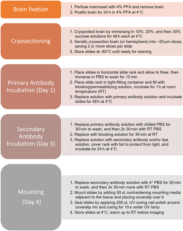
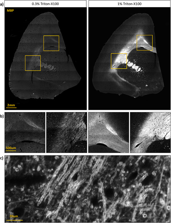
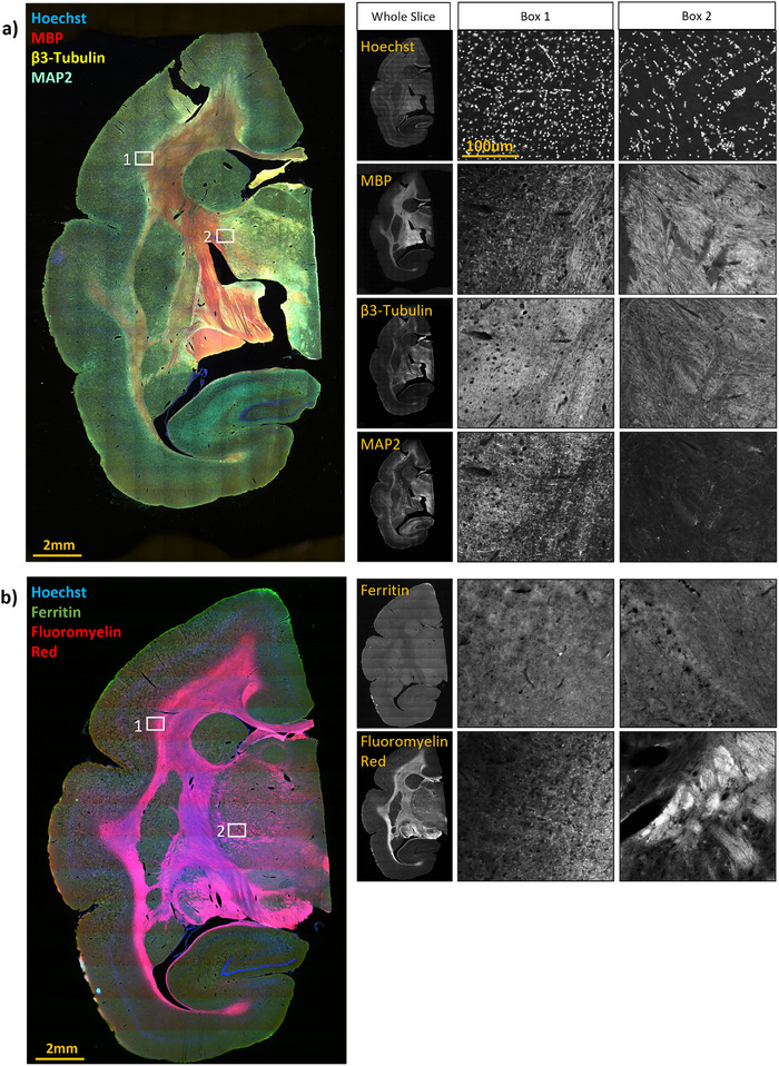
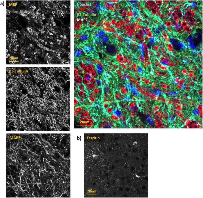
Here, we have characterized a set of marmoset-compatible fluorescent dyes and antibodies that label myelin, axons, dendrites, and the iron-storage protein ferritin, and developed a batch-style multiplex immunohistochemistry protocol to uniformly process large numbers of tissue slides for multiple cell-type specific markers.
We provide a practical guide for researchers interested in harnessing the potential of marmoset models to advance understanding of brain structure, function, and pathophysiology.
普通狨猴是一种小型非人灵长类动物,已成为神经科学研究中有价值的动物模型。对脑组织进行准确分析对于理解狨猴神经生理学以及模拟神经退行性疾病至关重要。迄今为止,许多研究已将磁共振成像(MRI)与组织化学染色相结合,而非免疫荧光标记,免疫荧光标记能够生成更具信息性和更高分辨率的图像。为新兴的狨猴研究领域生成资源,特别是用于展示不同细胞类型和其他结构组织的脑组织结构图谱,需要高通量免疫标记和成像方法。
在此,我们鉴定了一组与狨猴兼容的荧光染料和抗体,这些染料和抗体可标记髓磷脂、轴突、树突以及铁储存蛋白铁蛋白,并开发了一种批量式多重免疫组织化学方案,以统一处理大量用于多种细胞类型特异性标记的组织切片。
我们为有兴趣利用狨猴模型的潜力来增进对脑结构、功能和病理生理学理解的研究人员提供了一份实用指南。