Conze Christian, Trushina Nataliya I, Monteiro-Abreu Nanci, Singh Lisha, Romero Daniel Villar, Wienbeuker Eike, Schwarze Anna-Sophie, Holtmannspötter Michael, Bakota Lidia, Brandt Roland
Department of Neurobiology, School of Biology/Chemistry, Osnabrück University, Germany.
Center for Cellular Nanoanalytics, Osnabrück University, Germany.
Redox Biol. 2025 Jun;83:103626. doi: 10.1016/j.redox.2025.103626. Epub 2025 Apr 3.
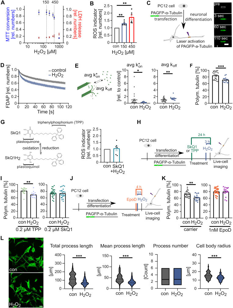
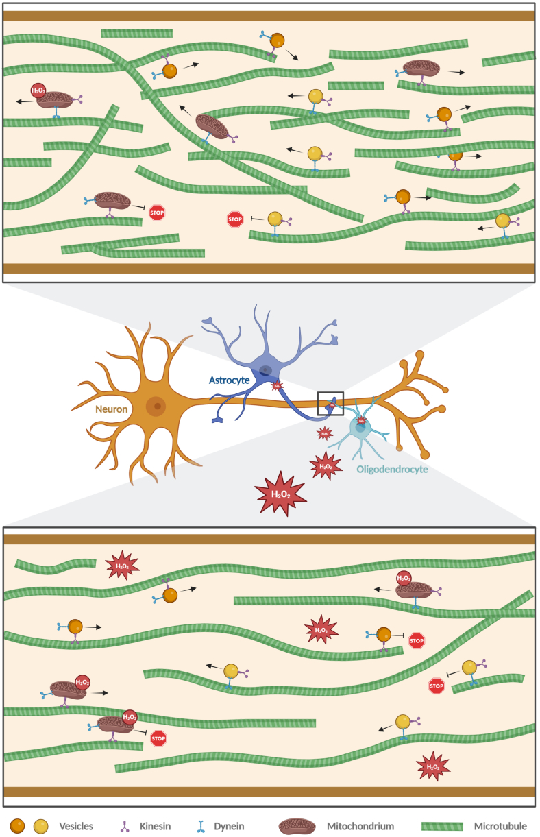
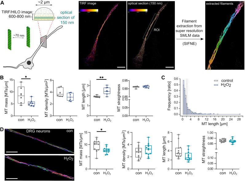
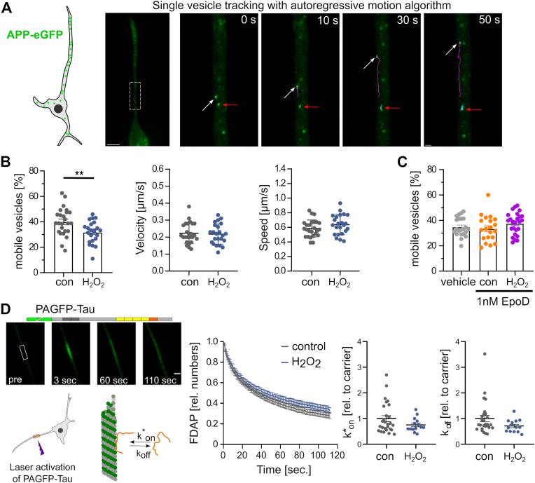
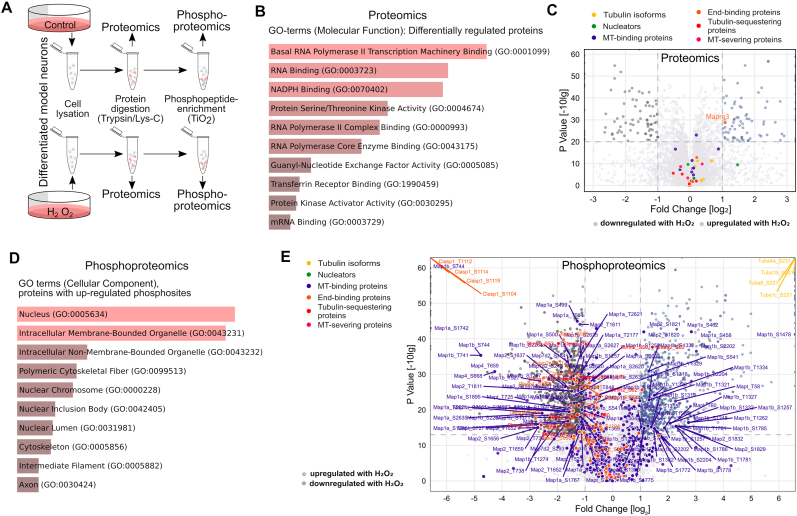
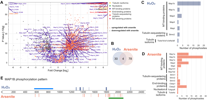
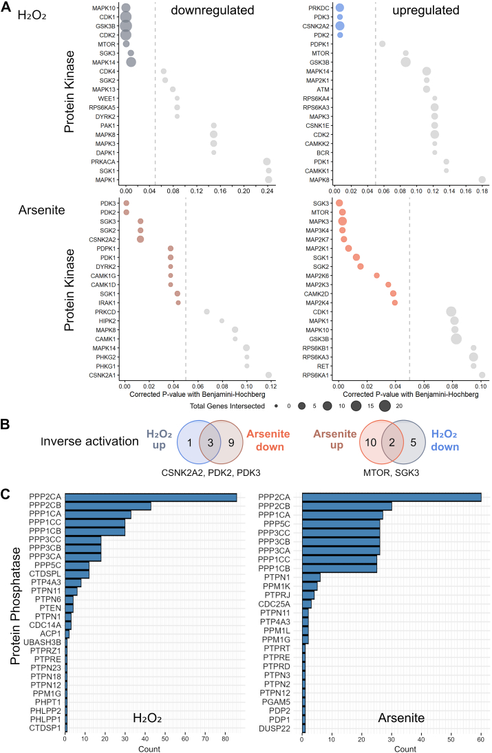
Many life processes are regulated by physiological redox signaling, but excessive oxidative stress can damage biomolecules and contribute to disease. Neuronal microtubules are critically involved in axon homeostasis, regulation of axonal transport, and neurodegenerative processes. However, whether and how physiological redox signaling affects axonal microtubules is largely unknown. Using live cell imaging and super-resolution microscopy, we show that subtoxic concentrations of the central redox metabolite hydrogen peroxide increase axonal microtubule dynamics, alter the structure of the axonal microtubule array, and affect the efficiency of axonal transport. We report that the mitochondria-targeting antioxidant SkQ1 and the microtubule stabilizer EpoD abolish the increase in microtubule dynamics. We found that hydrogen peroxide specifically modulates the phosphorylation state of microtubule-regulating proteins, which differs from arsenite as an alternative stress inducer, and induces a largely non-overlapping phosphorylation pattern of MAP1B as a main target. Cell-wide phosphoproteome analysis revealed signaling pathways that are inversely activated by hydrogen peroxide and arsenite. In particular, hydrogen peroxide treatment was associated with kinases that suppress apoptosis and regulate brain metabolism (PRKDC, CK2, PDKs), suggesting that these pathways play a central role in physiological redox signaling and modulation of axonal microtubule organization. The results suggest that the redox metabolite and second messenger hydrogen peroxide induces rapid and local reorganization of the microtubule array in response to mitochondrial activity or as a messenger from neighboring cells by activating specific signaling cascades.
许多生命过程受生理性氧化还原信号调控,但过度的氧化应激会损伤生物分子并导致疾病。神经元微管在轴突稳态、轴突运输调节及神经退行性过程中起关键作用。然而,生理性氧化还原信号是否以及如何影响轴突微管在很大程度上尚不清楚。利用活细胞成像和超分辨率显微镜技术,我们发现中心氧化还原代谢物过氧化氢的亚毒性浓度可增加轴突微管动力学,改变轴突微管阵列结构,并影响轴突运输效率。我们报道靶向线粒体的抗氧化剂SkQ1和微管稳定剂EpoD可消除微管动力学的增加。我们发现过氧化氢特异性调节微管调节蛋白的磷酸化状态,这与作为另一种应激诱导剂的亚砷酸盐不同,并诱导作为主要靶点的微管相关蛋白1B(MAP1B)产生很大程度上不重叠的磷酸化模式。全细胞磷酸化蛋白质组分析揭示了被过氧化氢和亚砷酸盐反向激活的信号通路。特别是,过氧化氢处理与抑制细胞凋亡和调节脑代谢的激酶(蛋白激酶DNA激活催化亚基、酪蛋白激酶2、丙酮酸脱氢酶激酶)相关,表明这些通路在生理性氧化还原信号及轴突微管组织调节中起核心作用。结果表明,氧化还原代谢物及第二信使过氧化氢通过激活特定信号级联反应,响应线粒体活性或作为来自相邻细胞的信使,诱导微管阵列的快速局部重组。