Hou Xuefeng, Chen Kun, Luo Huiwen, Xu Wengui, Li Xiaofeng
Department of Molecular Imaging and Nuclear Medicine, Tianjin Medical University Cancer Institute and Hospital, National Clinical Research Center for Cancer, Huan-Hu-Xi Road, Ti-Yuan-Bei, He Xi District, Tianjin, 300060, China.
Tianjin's Clinical Research Center for Cancer, Tianjin, 300060, China.
Cancer Imaging. 2025 May 12;25(1):62. doi: 10.1186/s40644-025-00880-2.
According to the updated classification system, human epidermal growth factor receptor 2 (HER2) expression statuses are divided into the following three groups: HER2-over-expression, HER2-low-expression, and HER2-zero-expression. HER2-negative expression was reclassified into HER2-low-expression and HER2-zero-expression. This study aimed to identify three different HER2 expression statuses for breast cancer (BC) patients using PET/CT radiomics and clinicopathological characteristics.
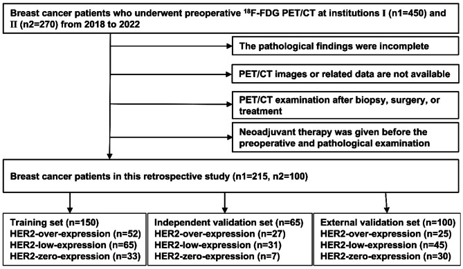
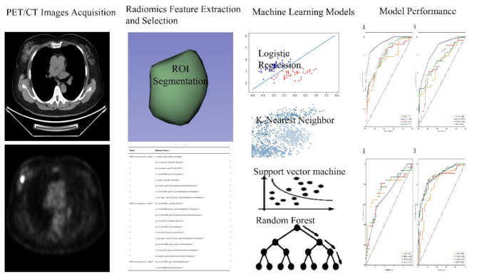
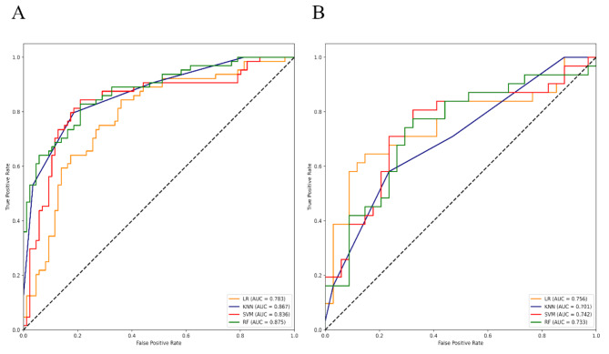
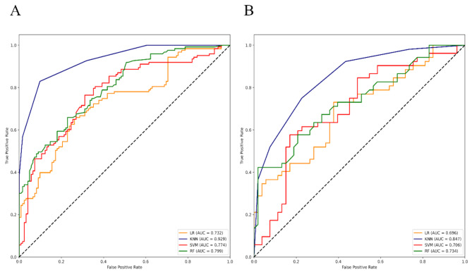
A total of 315 BC patients who met the inclusion and exclusion criteria from two institutions were retrospectively included. The patients in institution 1 were divided into the training set and the independent validation set according to the ratio of 7:3, and institution 2 was used as the external validation set. According to the results of pathological examination, all BC patients were divided into HER2-over-expression, HER2-low-expression, and HER2-zero-expression. First, PET/CT radiomic features and clinicopathological features based on each patient were extracted and collected. Second, multiple methods were used to perform feature screening and feature selection. Then, four machine learning classifiers, including logistic regression (LR), k-nearest neighbor (KNN), support vector machine (SVM), and random forest (RF), were constructed to identify HER2-over-expression vs. others, HER2-low-expression vs. others, and HER2-zero-expression vs. others. The receiver operator characteristic (ROC) curve was plotted to measure the model's predictive power.
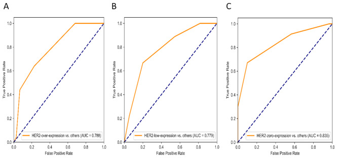
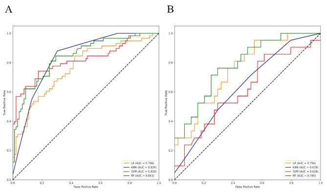
According to the feature screening process, 8, 10, and 2 radiomics features and 2 clinicopathological features were finally selected to construct three prediction models (HER2-over-expression vs. others, HER2-low-expression vs. others, and HER2-zero-expression vs. others). For HER2-over-expression vs. others, the RF model outperformed other models with an AUC value of 0.843 (95%CI: 0.774-0.897), 0.785 (95%CI: 0.665-0.877), and 0.788 (95%CI: 0.708-0.868) in the training set, independent validation set, and external validation set. Concerning HER2-low-expression vs. others, the outperformance of the LR model over other models was identified with an AUC value of 0.783 (95%CI: 0.708-0.846), 0.756 (95%CI: 0.634-0.854), and 0.779 (95%CI: 0.698-0.860) in the training set, independent validation set, and external validation set. Whereas, the KNN model was confirmed as the optimal model to distinguish HER2-zero-expression from others, with an AUC value of 0.929 (95%CI: 0.890-0.958), 0.847 (95%CI: 0.764-0.910), and 0.835 (95%CI: 0.762-0.908) in the training set, independent validation set, and external validation set.
Combined PET/CT radiomic models integrating with clinicopathological characteristics are non-invasively predictive of different HER2 statuses of BC patients.
根据更新后的分类系统,人表皮生长因子受体2(HER2)表达状态分为以下三组:HER2过表达、HER2低表达和HER2零表达。HER2阴性表达被重新分类为HER2低表达和HER2零表达。本研究旨在利用PET/CT影像组学和临床病理特征识别乳腺癌(BC)患者的三种不同HER2表达状态。
回顾性纳入来自两个机构的315例符合纳入和排除标准的BC患者。机构1中的患者按7:3的比例分为训练集和独立验证集,机构2用作外部验证集。根据病理检查结果,将所有BC患者分为HER2过表达、HER2低表达和HER2零表达。首先,提取并收集基于每位患者的PET/CT影像组学特征和临床病理特征。其次,使用多种方法进行特征筛选和特征选择。然后,构建包括逻辑回归(LR)、k近邻(KNN)、支持向量机(SVM)和随机森林(RF)在内的四个机器学习分类器,以识别HER2过表达与其他情况、HER2低表达与其他情况以及HER2零表达与其他情况。绘制受试者工作特征(ROC)曲线以衡量模型的预测能力。
根据特征筛选过程,最终选择8个、10个和2个影像组学特征以及2个临床病理特征来构建三个预测模型(HER2过表达与其他情况、HER2低表达与其他情况以及HER2零表达与其他情况)。对于HER2过表达与其他情况,RF模型在训练集、独立验证集和外部验证集中的AUC值分别为0.843(95%CI:0.774 - 0.897)、0.785(95%CI:0.665 - 0.877)和0.788(95%CI:0.708 - 0.868),优于其他模型。关于HER2低表达与其他情况,LR模型在训练集、独立验证集和外部验证集中的AUC值分别为0.783(95%CI:0.708 - 0.846)、0.756(95%CI:0.634 - 0.854)和0.779(95%CI:0.698 - 0.860),被确定优于其他模型。而KNN模型被确认为区分HER2零表达与其他情况的最佳模型,在训练集、独立验证集和外部验证集中的AUC值分别为0.929(95%CI:0.890 - 0.958)、0.847(95%CI:0.764 - 0.910)和0.835(95%CI:0.762 - 0.908)。
结合PET/CT影像组学模型与临床病理特征可对BC患者不同的HER2状态进行无创预测。