Xu Duo, Peng Tao, Cai Fuxin, Wang Lei, Chen Xinjian
School of Electronics and Information Engineering, Soochow University, Suzhou, China.
School of Future Science and Engineering, Soochow University, Suzhou, China.
Quant Imaging Med Surg. 2025 May 1;15(5):4311-4320. doi: 10.21037/qims-24-2051. Epub 2025 Apr 28.
As the use of ophthalmic optical biometers in clinical practice has increased, so too has the demand for higher standards in evaluating ocular metrics. Among these metrics, the measurement of the ocular axial length (AL) is a critical task that often requires calibrated devices. Thus, efficient algorithms need to be developed to improve calibration mechanisms and ensure precise measurements. This study aimed to establish an algorithm to determine the pixel heights (PHs) of the boundary vertices of a calibration device in ophthalmic optical biometers to improve the accuracy and repeatability of ocular AL measurements in optical coherence tomography (OCT) images.
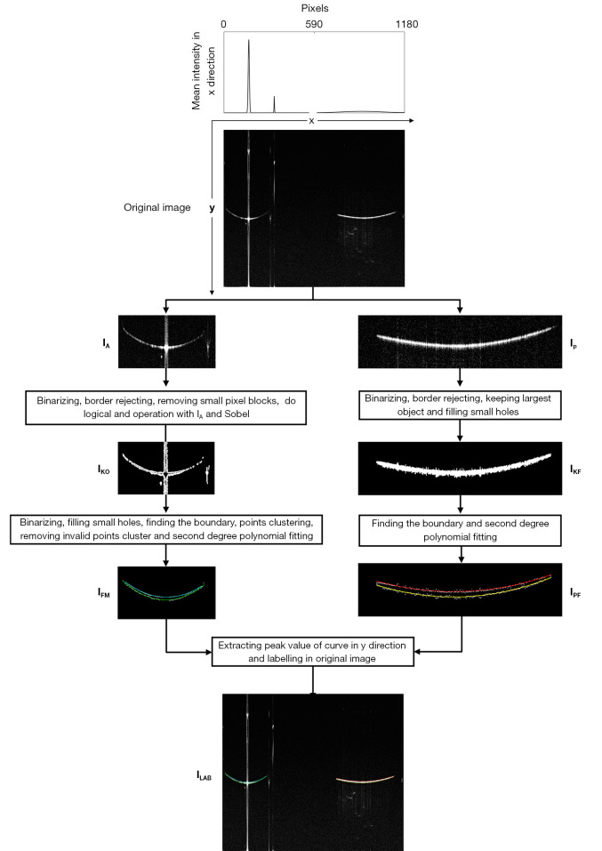
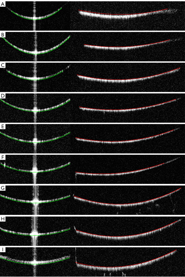
The algorithm employs a series of image morphological processing techniques to delineate the rough boundaries of the calibration device in OCT images. After extracting boundary points, clustering techniques are applied to simplify the data. These clustered boundary points are analyzed for characteristic parameters to refine the boundaries. Finally, a fitting process is used to determine the PHs of the vertices, and performance is then evaluated by tests measuring the repeatability and recognition accuracy of the algorithm.
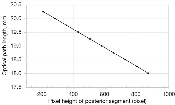
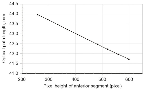
The proposed method had high repeatability in locating the boundaries of the front and rear sections of the calibration device, and had repeatability deviations not exceeding 0.36 pixels and 0.18 pixels, respectively. Additionally, linear fitting of the computed sub-PH and optical path difference yielded determination coefficients of 0.9998330 and 0.9999863, respectively, indicating an almost perfectly linear relationship. This high degree of linearity demonstrates the exceptional accuracy of the method in locating the calibration device boundaries. These results provide a heuristic approach for the future boundary localization of calibration devices in OCT images for biometers, offering a robust and reliable framework for enhancing calibration precision in ophthalmic optical biometry.
The developed algorithm provides an effective and reliable method for determining the PHs of calibration device vertices in ophthalmic optical biometers. With its high accuracy and repeatability, this valuable tool could enhance ocular AL measurements and improve the overall quality of ocular assessments in clinical practice.
随着眼科光学生物测量仪在临床实践中的应用增加,对评估眼部指标的更高标准的需求也随之增加。在这些指标中,眼轴长度(AL)的测量是一项关键任务,通常需要校准设备。因此,需要开发高效算法来改进校准机制并确保精确测量。本研究旨在建立一种算法,以确定眼科光学生物测量仪中校准设备边界顶点的像素高度(PH),从而提高光学相干断层扫描(OCT)图像中眼轴长度测量的准确性和可重复性。
该算法采用一系列图像形态处理技术来描绘OCT图像中校准设备的大致边界。提取边界点后,应用聚类技术简化数据。对这些聚类的边界点进行特征参数分析以细化边界。最后,使用拟合过程确定顶点的像素高度,然后通过测量算法的可重复性和识别准确性的测试来评估性能。
所提出的方法在校准设备前后部分边界定位方面具有高重复性,重复性偏差分别不超过0.36像素和0.18像素。此外,计算得到的子像素高度与光程差的线性拟合决定系数分别为0.9998330和0.9999863,表明几乎是完美的线性关系。这种高度的线性证明了该方法在定位校准设备边界方面的卓越准确性。这些结果为生物测量仪的OCT图像中校准设备的未来边界定位提供了一种启发式方法,为提高眼科光学生物测量中的校准精度提供了一个强大而可靠的框架。
所开发的算法为确定眼科光学生物测量仪中校准设备顶点的像素高度提供了一种有效且可靠的方法。凭借其高准确性和可重复性,这一有价值的工具可增强眼轴长度测量,并提高临床实践中眼部评估的整体质量。