• 文献检索
  • 文档翻译
  • 深度研究
  • 学术资讯
  • Suppr Zotero 插件Zotero 插件
  • 邀请有礼
  • 套餐&价格
  • 历史记录
应用&插件
Suppr Zotero 插件Zotero 插件浏览器插件Mac 客户端Windows 客户端微信小程序
定价
高级版会员购买积分包购买API积分包
服务
文献检索文档翻译深度研究API 文档MCP 服务
关于我们
关于 Suppr公司介绍联系我们用户协议隐私条款
关注我们

Suppr 超能文献

核心技术专利:CN118964589B侵权必究
粤ICP备2023148730 号-1Suppr @ 2026

文献检索

告别复杂PubMed语法,用中文像聊天一样搜索,搜遍4000万医学文献。AI智能推荐,让科研检索更轻松。

立即免费搜索

文件翻译

保留排版,准确专业,支持PDF/Word/PPT等文件格式,支持 12+语言互译。

免费翻译文档

深度研究

AI帮你快速写综述,25分钟生成高质量综述,智能提取关键信息,辅助科研写作。

立即免费体验

内视网膜变薄可作为首发帕金森病认知障碍的生物标志物。

Inner retinal thinning as a biomarker for cognitive impairment in de novo Parkinson's disease.

机构信息

Department of Ophthalmology, Chonnam National University Medical School and Hospital, Gwangju, South Korea.

Department of Neurology, Chonnam National University Medical School and Hospital, Gwangju, South Korea.

出版信息

Sci Rep. 2019 Aug 14;9(1):11832. doi: 10.1038/s41598-019-48388-7.

DOI:10.1038/s41598-019-48388-7
PMID:31413302
原文链接:https://pmc.ncbi.nlm.nih.gov/articles/PMC6694167/
Abstract

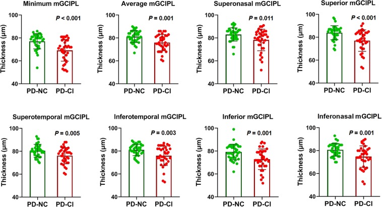
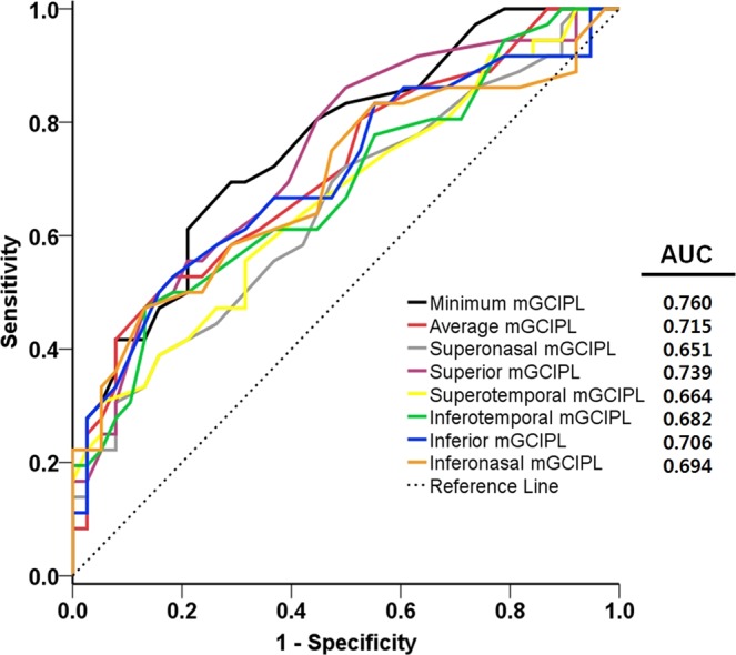
We investigated the association between retinal changes measured using optical coherence tomography (OCT) and diverse clinical grading scales in patients with Parkinson's disease (PD). Seventy-four eyes of 74 patients with de novo PD and 53 eyes of age-matched control subjects were included. The thickness of the peripapillary retinal nerve fiber layer (pRNFL) and macular ganglion cell-inner plexiform layer (mGCIPL) were measured. We analyzed the correlations between the clinical PD grading scales and OCT parameters, and between the OCT parameters and volumetric data in the cerebral cortical and subcortical structures. The area under the receiver operating characteristic curve (AUC) was calculated for diagnosing cognitive impairment in patients with PD. Statistically significant reductions in the thickness of average, temporal, and inferior pRNFL and overall mGCIPL were observed in patients with PD. The Montreal Cognitive Assessment score was significantly associated with mGCIPL thinning. The AUC of the mGCIPL parameters for diagnosing cognitive impairment in patients with PD ranged from 0.651 to 0.760. Moreover, thinning of the mGCIPL was significantly associated with the volumetric parameters of associated brain structures. Our findings highlight the clinical implications of OCT measurements as a potential biomarker for early detection of cognitive impairment in patients with PD.

摘要

我们研究了使用光学相干断层扫描(OCT)测量的视网膜变化与帕金森病(PD)患者多种临床分级量表之间的关系。纳入了 74 例初诊 PD 患者的 74 只眼和 53 只年龄匹配的对照者的眼。测量了视盘周围视网膜神经纤维层(pRNFL)和黄斑神经节细胞-内丛状层(mGCIPL)的厚度。我们分析了临床 PD 分级量表与 OCT 参数之间、OCT 参数与大脑皮质和皮质下结构容积数据之间的相关性。计算了 OCT 诊断 PD 患者认知障碍的受试者工作特征曲线(ROC)下面积(AUC)。PD 患者的平均、颞侧和下侧 pRNFL 以及整体 mGCIPL 厚度明显降低。蒙特利尔认知评估评分与 mGCIPL 变薄显著相关。mGCIPL 参数诊断 PD 患者认知障碍的 AUC 范围为 0.651 至 0.760。此外,mGCIPL 变薄与相关脑结构的容积参数显著相关。我们的研究结果强调了 OCT 测量作为早期检测 PD 患者认知障碍的潜在生物标志物的临床意义。

https://cdn.ncbi.nlm.nih.gov/pmc/blobs/d4e9/6694167/055dfed157b4/41598_2019_48388_Fig2_HTML.jpg
https://cdn.ncbi.nlm.nih.gov/pmc/blobs/d4e9/6694167/dec949f01409/41598_2019_48388_Fig1_HTML.jpg
https://cdn.ncbi.nlm.nih.gov/pmc/blobs/d4e9/6694167/055dfed157b4/41598_2019_48388_Fig2_HTML.jpg
https://cdn.ncbi.nlm.nih.gov/pmc/blobs/d4e9/6694167/dec949f01409/41598_2019_48388_Fig1_HTML.jpg
https://cdn.ncbi.nlm.nih.gov/pmc/blobs/d4e9/6694167/055dfed157b4/41598_2019_48388_Fig2_HTML.jpg

相似文献

1
Inner retinal thinning as a biomarker for cognitive impairment in de novo Parkinson's disease.内视网膜变薄可作为首发帕金森病认知障碍的生物标志物。
Sci Rep. 2019 Aug 14;9(1):11832. doi: 10.1038/s41598-019-48388-7.
2
Temporal Relation between Macular Ganglion Cell-Inner Plexiform Layer Loss and Peripapillary Retinal Nerve Fiber Layer Loss in Glaucoma.青光眼的黄斑神经节细胞-内丛状层丢失与视盘周围视网膜神经纤维层丢失的时间关系。
Ophthalmology. 2017 Jul;124(7):1056-1064. doi: 10.1016/j.ophtha.2017.03.014. Epub 2017 Apr 10.
3
Diagnostic ability of macular ganglion cell inner plexiform layer measurements in glaucoma using swept source and spectral domain optical coherence tomography.使用扫频源和谱域光学相干断层扫描测量黄斑神经节细胞内丛状层在青光眼诊断中的能力
PLoS One. 2015 May 15;10(5):e0125957. doi: 10.1371/journal.pone.0125957. eCollection 2015.
4
Retinal and Choroidal Changes in Patients with Parkinson's Disease Detected by Swept-Source Optical Coherence Tomography.扫频光学相干断层扫描检测帕金森病患者的视网膜和脉络膜变化
Curr Eye Res. 2018 Jan;43(1):109-115. doi: 10.1080/02713683.2017.1370116. Epub 2017 Nov 7.
5
Macular inner plexiform and retinal nerve fiber layer thickness in glaucoma.青光眼患者黄斑内丛状层及视网膜神经纤维层厚度
Optom Vis Sci. 2014 Nov;91(11):1320-7. doi: 10.1097/OPX.0000000000000392.
6
Macular Ganglion Cell-Inner Plexiform Layer Loss Precedes Peripapillary Retinal Nerve Fiber Layer Loss in Glaucoma with Lower Intraocular Pressure.在眼压较低的青光眼患者中,黄斑神经节细胞-内丛状层损失先于视盘周围视网膜神经纤维层损失。
Ophthalmology. 2019 Aug;126(8):1119-1130. doi: 10.1016/j.ophtha.2019.03.016. Epub 2019 Mar 22.
7
Early macular ganglion cell-inner plexiform layer analysis in non-arteritic anterior ischemic optic neuropathy.非动脉炎性前部缺血性视神经病变的早期黄斑神经节细胞-内层神经丛层分析
Graefes Arch Clin Exp Ophthalmol. 2016 May;254(5):983-9. doi: 10.1007/s00417-015-3230-9. Epub 2015 Dec 3.
8
Temporal Raphe Sign for Discrimination of Glaucoma from Optic Neuropathy in Eyes with Macular Ganglion Cell-Inner Plexiform Layer Thinning.时间隔核标志可用于鉴别伴黄斑神经节细胞-内丛状层变薄的青光眼和视神经病变
Ophthalmology. 2019 Aug;126(8):1131-1139. doi: 10.1016/j.ophtha.2018.12.031. Epub 2018 Dec 18.
9
Parafoveal thinning of inner retina is associated with visual dysfunction in Lewy body diseases.周边部视网膜内层变薄与路易体病的视觉功能障碍有关。
Mov Disord. 2019 Sep;34(9):1315-1324. doi: 10.1002/mds.27728. Epub 2019 May 28.
10
Ganglion Cell-Inner Plexiform Layer and Peripapillary Retinal Nerve Fiber Layer Thicknesses in Age-Related Macular Degeneration.年龄相关性黄斑变性中神经节细胞-内网状层及视乳头周围视网膜神经纤维层厚度
Invest Ophthalmol Vis Sci. 2015 Jun;56(6):3976-83. doi: 10.1167/iovs.15-17013.

引用本文的文献

1
Visualizing the invisible: inner plexiform layer stratification with conventional spectral-domain optical coherence tomography.可视化不可见之物:利用传统光谱域光学相干断层扫描技术对内核层进行分层
Int J Retina Vitreous. 2025 Jun 15;11(1):65. doi: 10.1186/s40942-025-00692-3.
2
Retinal Thickness in Patients with Parkinson's Disease and Dopa Responsive Dystonia-Is There Any Difference?帕金森病和多巴反应性肌张力障碍患者的视网膜厚度——有差异吗?
Biomedicines. 2025 May 19;13(5):1227. doi: 10.3390/biomedicines13051227.
3
Cognition-Associated Changes in Retinal Thickness Relate to Limbic and Temporal Cortical Atrophy in Parkinson's Disease.

本文引用的文献

1
Retinal α-synuclein deposits in Parkinson's disease patients and animal models.帕金森病患者和动物模型中的视网膜 α-突触核蛋白沉积。
Acta Neuropathol. 2019 Mar;137(3):379-395. doi: 10.1007/s00401-018-01956-z. Epub 2019 Feb 5.
2
Higher serum triglyceride levels are associated with Parkinson's disease mild cognitive impairment.较高的血清甘油三酯水平与帕金森病轻度认知障碍有关。
Mov Disord. 2018 Dec;33(12):1970-1971. doi: 10.1002/mds.27521. Epub 2018 Oct 21.
3
Association of Retinal Neurodegeneration on Optical Coherence Tomography With Dementia: A Population-Based Study.
帕金森病中视网膜厚度与认知相关的变化与边缘系统和颞叶皮质萎缩有关。
Brain Behav. 2025 May;15(5):e70509. doi: 10.1002/brb3.70509.
4
Idiopathic Parkinson's Disease and Neuro-ophthalmological Findings: A Study on Neurodegeneration in the Retinal Nerve Fiber Layer and Cognitive Functions.特发性帕金森病与神经眼科检查结果:视网膜神经纤维层神经退行性变及认知功能的研究
Noro Psikiyatr Ars. 2025 Feb 20;62(1):77-83. doi: 10.29399/npa.28670. eCollection 2025.
5
From Diagnosis to Treatment: A Comprehensive Review of Biomarkers and Therapeutic Advances in Parkinson's Disease.从诊断到治疗:帕金森病生物标志物与治疗进展的全面综述
Ann Neurosci. 2025 Jan;32(1):51-57. doi: 10.1177/09727531231200733. Epub 2023 Nov 1.
6
Dynamics of retinal changes in early-stage Parkinson's disease.早期帕金森病视网膜变化的动态研究
Acta Neuropathol Commun. 2025 Jan 31;13(1):20. doi: 10.1186/s40478-025-01936-x.
7
Macular structural integrity estimates are associated with Parkinson's disease genetic risk.黄斑结构完整性估计与帕金森病遗传风险相关。
Acta Neuropathol Commun. 2024 Aug 13;12(1):130. doi: 10.1186/s40478-024-01841-9.
8
Neuroretinal Alterations in Schizophrenia and Bipolar Disorder: An Updated Meta-analysis.精神分裂症和双相情感障碍的神经视网膜改变:一项更新的荟萃分析。
Schizophr Bull. 2024 Aug 27;50(5):1067-1082. doi: 10.1093/schbul/sbae102.
9
Reduced Thickness of the Retina in de novo Parkinson's Disease Shows A Distinct Pattern, Different from Glaucoma.初发帕金森病患者的视网膜变薄呈现出一种独特的模式,与青光眼不同。
J Parkinsons Dis. 2024;14(3):507-519. doi: 10.3233/JPD-223481.
10
Association of retinal neurodegeneration with the progression of cognitive decline in Parkinson's disease.视网膜神经变性与帕金森病认知功能减退进展的关联
NPJ Parkinsons Dis. 2024 Jan 23;10(1):26. doi: 10.1038/s41531-024-00637-x.
基于人群的研究:光学相干断层扫描视网膜神经退行性变与痴呆的相关性。
JAMA Neurol. 2018 Oct 1;75(10):1256-1263. doi: 10.1001/jamaneurol.2018.1563.
4
Outer Retinal Assessment Using Spectral-Domain Optical Coherence Tomography in Patients With Alzheimer's and Parkinson's Disease.使用谱域光学相干断层扫描评估阿尔茨海默病和帕金森病患者的视网膜外层。
Invest Ophthalmol Vis Sci. 2018 Jun 1;59(7):2768-2777. doi: 10.1167/iovs.17-23240.
5
Volumetric brain analysis as a predictor of a worse cognitive outcome in Parkinson's disease.容积脑分析可预测帕金森病认知结局更差。
J Psychiatr Res. 2018 Jul;102:254-260. doi: 10.1016/j.jpsychires.2018.04.016. Epub 2018 Apr 27.
6
Apolipoprotein Eε4: A Biomarker for Executive Dysfunction among Parkinson's Disease Patients with Mild Cognitive Impairment.载脂蛋白Eε4:轻度认知障碍帕金森病患者执行功能障碍的生物标志物。
Front Neurosci. 2017 Dec 20;11:712. doi: 10.3389/fnins.2017.00712. eCollection 2017.
7
Patterns of Retinal Ganglion Cell Damage in Neurodegenerative Disorders: Parvocellular vs Magnocellular Degeneration in Optical Coherence Tomography Studies.神经退行性疾病中视网膜神经节细胞损伤模式:光学相干断层扫描研究中的小细胞与大细胞变性
Front Neurol. 2017 Dec 22;8:710. doi: 10.3389/fneur.2017.00710. eCollection 2017.
8
Imaging biomarkers in Parkinson's disease and Parkinsonian syndromes: current and emerging concepts.帕金森病及帕金森综合征中的影像学生物标志物:当前概念与新进展
Transl Neurodegener. 2017 Mar 28;6:8. doi: 10.1186/s40035-017-0076-6. eCollection 2017.
9
In Vivo Morphology of the Optic Nerve and Retina in Patients With Parkinson's Disease.帕金森病患者视神经和视网膜的体内形态学
Invest Ophthalmol Vis Sci. 2016 Aug 1;57(10):4420-4427. doi: 10.1167/iovs.16-20020.
10
Clinical Usefulness of Spectral-Domain Optical Coherence Tomography in Glaucoma and NAION.频域光学相干断层扫描在青光眼和非动脉炎性前部缺血性视神经病变中的临床应用价值
Chonnam Med J. 2016 Sep;52(3):194-200. doi: 10.4068/cmj.2016.52.3.194. Epub 2016 Sep 23.