Xu Zhirong, Ye Jiayi, Wang Han, Chen Jiemin, Tan Kailing, Li Shilin, Su Shanshan
Department of Ultrasound, The Second Affiliated Hospital of Fujian Medical University, Quanzhou 362000, China.
Department of Nuclear Medicine, The Second Affiliated Hospital of Fujian Medical University, Quanzhou 362000, China.
Diagnostics (Basel). 2025 Jun 15;15(12):1519. doi: 10.3390/diagnostics15121519.
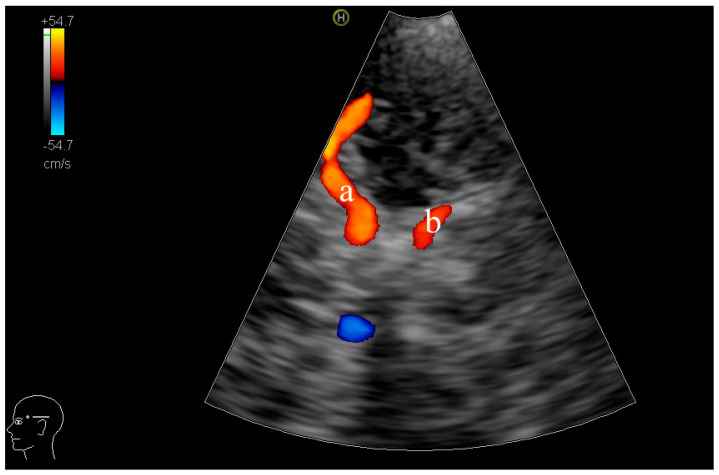
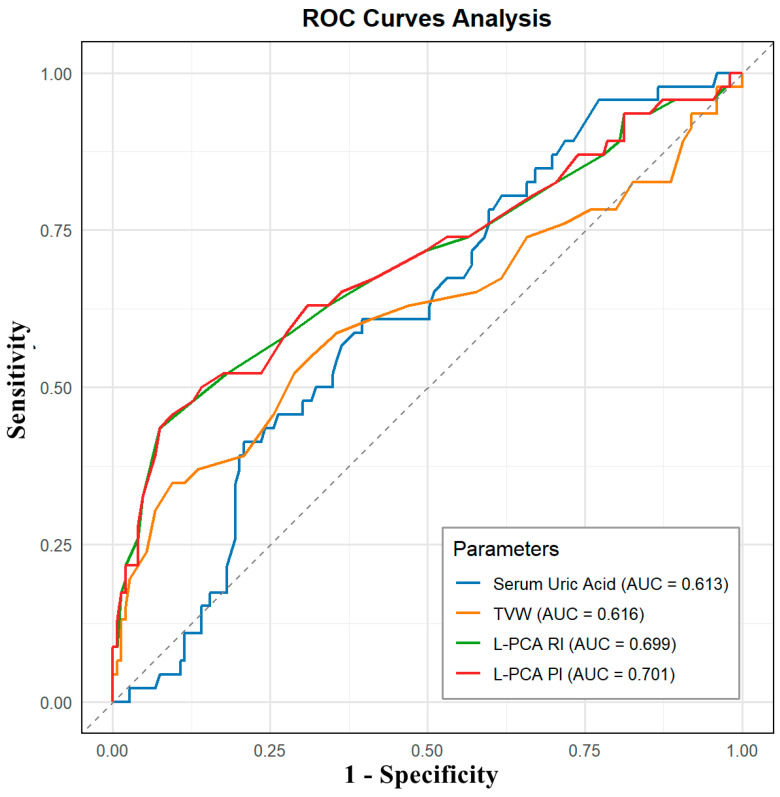
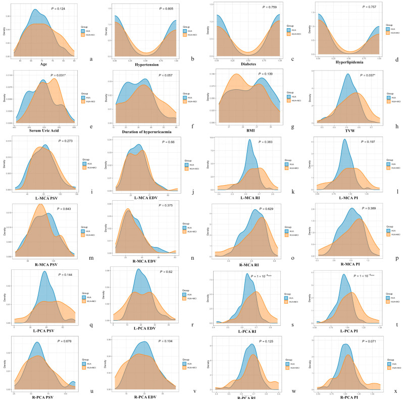
Hyperuricaemia has been linked to cognitive decline, yet cerebral structural and haemodynamic changes in this population remain poorly defined. We evaluated transcranial colour-coded duplex (TCCD) sonography as a non-invasive screening tool for early mild cognitive impairment (MCI) in elderly hyperuricaemic men. In this cross-sectional study, 195 men aged ≥ 60 years with hyperuricaemia were stratified by the Montreal Cognitive Assessment (MoCA) into HUA + MCI (MoCA < 26, = 46) and HUA (MoCA ≥ 26, = 149) groups. TCCD measured third-ventricle width (TVW) and peak systolic/end-diastolic velocities to calculate resistive (RI) and pulsatility (PI) indices in the middle (MCA) and posterior (PCA) cerebral arteries. Serum uric acid was recorded. Kernel density plots and receiver operating characteristic (ROC) curves assessed diagnostic performance. The HUA + MCI group exhibited higher serum uric acid (508.5 ± 36.3 vs. 492.9 ± 44.0 µmol/L; = 0.031), greater TVW (0.55 ± 0.11 vs. 0.51 ± 0.08 cm; = 0.037), and elevated left PCA RI (0.69 ± 0.07 vs. 0.64 ± 0.06) and PI (1.05 ± 0.17 vs. 0.95 ± 0.12; both < 0.001). ROC analysis identified left PCA PI as the most specific marker (AUC = 0.701; specificity 90.6%; sensitivity 45.7%). Kernel density plots confirmed distinct distributions of key parameters. TCCD-detected ventricular enlargement and raised PCA pulsatility accurately distinguish MCI among hyperuricaemic men. As a non-invasive, accessible technique with high specificity, TCCD may complement MRI and cognitive testing in early screening of at-risk populations.
高尿酸血症与认知功能下降有关,但该人群的脑结构和血流动力学变化仍不清楚。我们评估了经颅彩色编码双功(TCCD)超声检查作为老年高尿酸血症男性早期轻度认知障碍(MCI)的一种非侵入性筛查工具。在这项横断面研究中,195名年龄≥60岁的高尿酸血症男性根据蒙特利尔认知评估(MoCA)分为高尿酸血症合并MCI组(MoCA<26,n = 46)和高尿酸血症组(MoCA≥26,n = 149)。TCCD测量第三脑室宽度(TVW)以及收缩期峰值/舒张末期速度,以计算大脑中动脉(MCA)和大脑后动脉(PCA)的阻力指数(RI)和搏动指数(PI)。记录血清尿酸水平。核密度图和受试者工作特征(ROC)曲线评估诊断性能。高尿酸血症合并MCI组的血清尿酸水平更高(508.5±36.3 vs. 492.9±44.0 µmol/L;P = 0.031),TVW更大(0.55±0.11 vs. 0.51±0.08 cm;P = 0.037),左PCA的RI(0.69±0.07 vs. 0.64±0.06)和PI升高(1.05±0.17 vs. 0.95±0.12;均P<0.001)。ROC分析确定左PCA的PI是最具特异性的标志物(AUC = 0.701;特异性90.6%;敏感性45.7%)。核密度图证实了关键参数的不同分布。TCCD检测到的脑室扩大和PCA搏动增加可准确区分高尿酸血症男性中的MCI。作为一种具有高特异性的非侵入性、可及性技术,TCCD可在早期筛查高危人群中补充MRI和认知测试。