Clodi Nikolaus, Bender Benjamin, Hecke Gretha, Hauptvogel Karolin, Gohla Georg, Hauser Till-Karsten, Ghibes Patrick, Hergan Klaus, Ernemann Ulrike, Estler Arne
Department of Radiology, Paracelsus Medical University of Salzburg, 5020 Salzburg, Austria.
Department of Diagnostic and Interventional Neuroradiology, University Hospital Tübingen, 72076 Tübingen, Germany.
Diagnostics (Basel). 2025 Jun 16;15(12):1523. doi: 10.3390/diagnostics15121523.
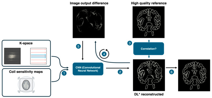
Assessing hippocampal pathology in epilepsy is challenging, and improving diagnostic accuracy can benefit from deep learning image reconstruction, standardized imaging protocols, and advanced post-processing methods. This study compares T2 TSE DRB (Deep Resolve Boost) sequences with standard T2 TSE sequences for hippocampal segmentation and volumetry using FreeSurfer, focusing on how DRB affects image acquisition time without compromising diagnostic accuracy. FreeSurfer (version 7.4.1) was used to segment hippocampal subregions in 36 subjects (mean age of 39 ± 14 years; 21 males, 15 females) using both T2 TSE DRB and T2 TSE sequences. The segmented volumes were compared with a two-tailed -test, and pathological volume differences were assessed using z-values based on a 95% confidence interval (-2 < z < 2). Overall hippocampal segment volumes were identical between sequences. However, significant volume differences were noted in the CA1-Body ( = 0.003), CA4-Body ( = 0.002), and whole hippocampal body ( = 0.012) in the right hippocampus. Despite these differences, the low effect sizes suggest DRB sequences are comparable to conventional sequences. Additionally, DRB reduced image acquisition time by 61%. Z-scores identified pathological volume changes between the left and right hippocampus in individual subjects. T2 TSE DRB sequences are non-inferior to conventional T2 TSE sequences for hippocampal segmentation. The DRB method improves efficiency while providing clinically reliable results, and the proposed 95% confidence interval can aid in more objective assessments of hippocampal pathology.
评估癫痫中的海马病理具有挑战性,而提高诊断准确性可受益于深度学习图像重建、标准化成像协议和先进的后处理方法。本研究使用FreeSurfer将T2 TSE DRB(深度解析增强)序列与标准T2 TSE序列进行比较,用于海马分割和容积测量,重点关注DRB如何在不影响诊断准确性的情况下影响图像采集时间。使用FreeSurfer(版本7.4.1)对36名受试者(平均年龄39±14岁;21名男性,15名女性)的海马亚区域进行分割,同时使用T2 TSE DRB和T2 TSE序列。将分割后的容积进行双尾检验比较,并使用基于95%置信区间(-2 < z < 2)的z值评估病理容积差异。序列间总体海马分割容积相同。然而,右侧海马的CA1-体部(P = 0.003)、CA4-体部(P = 0.002)和整个海马体部(P = 0.012)存在显著的容积差异。尽管存在这些差异,但低效应量表明DRB序列与传统序列相当。此外,DRB将图像采集时间减少了61%。Z分数确定了个体受试者左右海马之间的病理容积变化。对于海马分割,T2 TSE DRB序列不劣于传统的T2 TSE序列。DRB方法在提高效率的同时提供了临床可靠的结果,并且所提出的95%置信区间有助于更客观地评估海马病理。