Martel Anne-Caroline, Pittard Damien, Devergnas Annaelle, Risk Benjamin, Nassi Jonathan J, Yu Waylin, Downer Joshua D, Wichmann Thomas, Galvan Adriana
Emory National Primate Research Center, Emory University, Atlanta, GA 30329, USA.
Udall Center of Excellence for Parkinson's Disease Research, Emory University, Atlanta, GA 30329, USA.
iScience. 2025 May 27;28(6):112767. doi: 10.1016/j.isci.2025.112767. eCollection 2025 Jun 20.
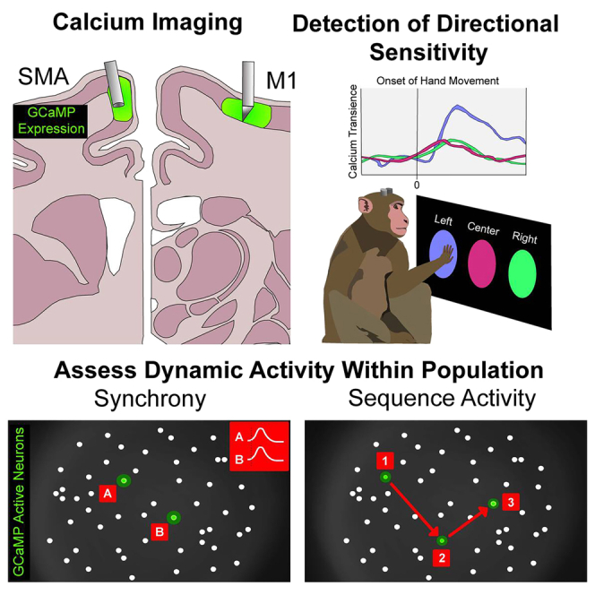
The study of motor cortices in non-human primates is relevant to our understanding of human motor control, both in healthy conditions and in movement disorders. Calcium imaging and miniature microscopes allow the study of multiple genetically identified neurons with excellent spatial resolution. We used this method to examine activity patterns of projection neurons in deep layers of the supplementary motor (SMA) and primary motor areas (M1) in four rhesus macaques. We implanted gradient index lenses and expressed GCaMP6f to image calcium transients while the animals were at rest or engaged in an arm-reaching task. We tracked the activity of SMA and M1 neurons across conditions, examined cell pairs for synchronous activity, and assessed whether SMA and M1 neuronal activation followed specific sequential activation patterns. We demonstrate the value of calcium imaging for studying patterns of activity in groups of corticofugal neurons in SMA and M1.
对非人类灵长类动物运动皮层的研究,对于我们理解健康状态以及运动障碍情况下的人类运动控制都具有重要意义。钙成像和微型显微镜能够以出色的空间分辨率对多个经基因鉴定的神经元进行研究。我们运用这种方法,对四只恒河猴的辅助运动区(SMA)和初级运动区(M1)深层的投射神经元的活动模式进行了检测。我们植入了梯度折射率透镜并表达了GCaMP6f,以便在动物休息或进行伸臂任务时对钙瞬变进行成像。我们追踪了不同条件下SMA和M1神经元的活动,检查细胞对的同步活动,并评估SMA和M1神经元激活是否遵循特定的顺序激活模式。我们证明了钙成像在研究SMA和M1中皮质传出神经元群活动模式方面的价值。