Rojas-Rueda Silvia, Watanabe Hidehiko, Abuhammoud Salah, Jurado Carlos A, Alshehri Abdullah, Fu Chin-Chuan, Vegh Daniel, Aldosary Khalid M, Algamaiah Hamad, Alshabib Abdulrahman
Resident, Master of Science in Dental Biomaterials, University of Alabama at Birmingham School of Dentistry, Birmingham, AL, USA.
Department of Oral Rehabilitation and Biosciences, Oregon Health & Sciences University School of Dentistry, Portland, OR, USA.
Saudi Dent J. 2025 Jul 1;37(4-6):27. doi: 10.1007/s44445-025-00029-8.
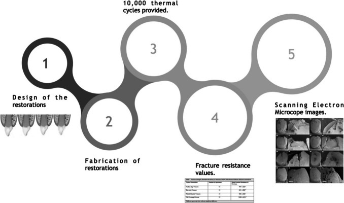
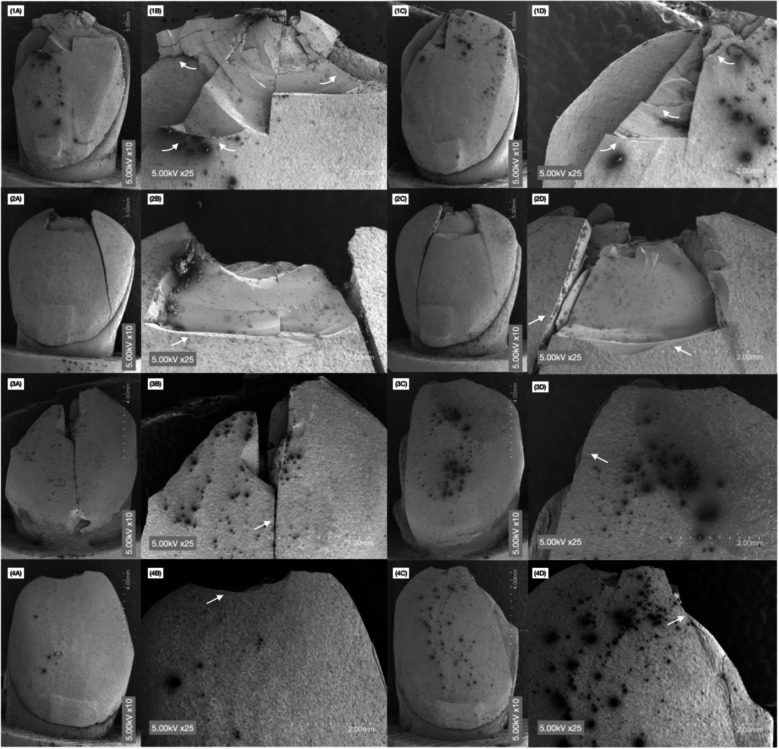
This study aimed to evaluate the fracture resistance of maxillary veneers with feather edge, butt-joint and palatal chamfer and traditional full coverage crowns fabricated out of chairside CAD/CAM advanced lithium disilicate and virgilite. Fifty-two restorations for maxillary right canine were fabricated (n = 13 per group) as follows: veneers with feather edge, veneers with butt-joint, veneers with palatal chamfer and full coverage crowns out of chairside CAD/CAM lithium disilicate and virgilite (Cerec Tessera). The restorations were bonded to 3D printed resin dies with resin cement (Variolink Esthetic LC). The cemented restorations were subjected to 10,000 thermocycles at 5 to 55 °C with a dwell time of 30 s. The specimens were loaded until fracture using a universal testing machine and the resistance was recorded in Newtons. Two-way ANOVA was used to assess the fracture resistance among veneers with different incisal edge designs and between veneers and crowns. Scanning electron microscope (SEM) images of the fractured specimens were taken and descriptive analysis was carried out. Full coverage crowns displayed higher fracture resistance (1496 ± 41 N) than any type of veneers. Veneers with palatal chamfer showed the highest value (842 ± 28 N) among veneers followed by butt joint veneers (661 ± 22 N). Feather edge veneers provided the lowest fracture resistance values (464 ± 23 N). The fracture resistance of CAD/CAM advanced lithium disilicate maxillary veneers are significantly influenced by the incisal edge design. Palatal chamfer veneers displayed higher fracture resistance than feather edge and butt joint veneers. Full coverage crowns offered higher fracture resistance than any type of veneer.
本研究旨在评估采用椅旁计算机辅助设计/计算机辅助制造(CAD/CAM)的高级二硅酸锂和碧玺制作的、具有羽状边缘、对接和腭侧倒角的上颌贴面以及传统全冠的抗折性。制作了52个上颌右侧尖牙修复体(每组n = 13),具体如下:羽状边缘贴面、对接贴面、腭侧倒角贴面以及采用椅旁CAD/CAM二硅酸锂和碧玺(Cerec Tessera)制作的全冠。使用树脂粘结剂(Variolink Esthetic LC)将修复体粘结到3D打印的树脂代型上。将粘结后的修复体在5至55°C下进行10000次热循环,保压时间为30秒。使用万能试验机对试件加载直至断裂,并记录以牛顿为单位的抗力。采用双向方差分析评估不同切缘设计的贴面之间以及贴面与全冠之间的抗折性。拍摄断裂试件的扫描电子显微镜(SEM)图像并进行描述性分析。全冠显示出比任何类型的贴面都更高的抗折性(1496±41 N)。在贴面中,腭侧倒角贴面显示出最高值(842±28 N),其次是对接贴面(661±22 N)。羽状边缘贴面提供的抗折性值最低(464±23 N)。CAD/CAM高级二硅酸锂上颌贴面的抗折性受切缘设计的显著影响。腭侧倒角贴面显示出比羽状边缘和对接贴面更高的抗折性。全冠提供的抗折性比任何类型的贴面都更高。