Gasteau Damien, Vrignaud Alexis, Biallais Arnaud, Richard Fabrice, Blancho Gilles, Branchereau Julien, Mesnard Benoît
DeepColor Imaging SAS, Nantes, France.
Nantes Université, CHU Nantes1, INSERM, Centre for Research in Transplantation and Translational Immunology, Nantes, France.
Eur Radiol Exp. 2025 Jul 9;9(1):65. doi: 10.1186/s41747-025-00601-1.
To evaluate in vivo a fully integrated photoacoustic tomography imaging system based on Fabry-Pérot ultrasound sensing method applied on porcine abdominal organs. This approach could be used by surgeons during intraoperative clinical procedures.
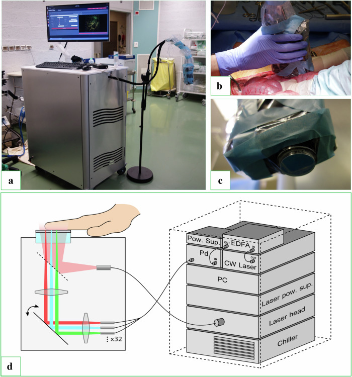
The photoacoustic imaging system was fully integrated into a single structure, and the detection technology was based on a Fabry-Pérot interferometer. The detection probe connected to the imaging system was applied directly to the organs of a male "large white" Sus scrofa pig weighing 80 kg, either manually or using a stand, with or without a gel interface. All experiments were performed in compliance with EU Directive 2010/63/EU on animal experimentation (APAFiS #31507).
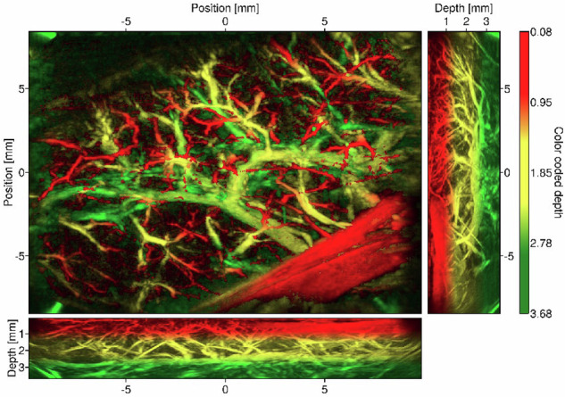
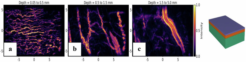
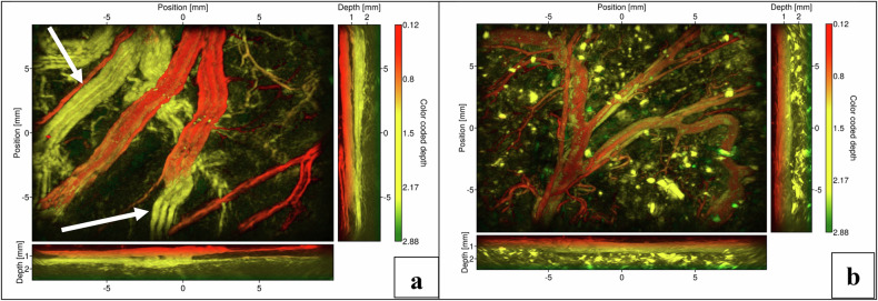
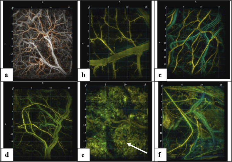
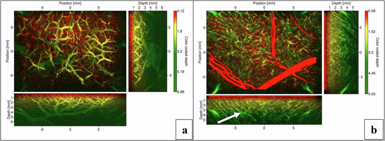
All intraperitoneal and retroperitoneal organs were evaluated using photoacoustic imaging. The evaluation of both hollow and solid organs was successfully conducted with consistent three-dimensional image quality. We demonstrate the system's ability to image blood vessels with diameters ranging from several millimeters down to less than 100 µm. Macroscopic evaluation of the organs using photoacoustic tomography imaging did not reveal any damage or burns caused by the excitation laser.
To our knowledge, this is the first reported imaging session of abdominal organs in an in vivo porcine model, performed using a photoacoustic tomography system with Fabry-Pérot interferometer detection. We present a high-resolution photoacoustic tomography system that is closer to routine clinical translation, thanks to a fully integrated system.
Photoacoustic evaluation of organs using a fully integrated system could become a valuable tool for surgical teams for intraprocedural assessment of vascularization.
Photoacoustic imaging visualizes blood vessels without contrast agents or ionizing radiation. Photoacoustic imaging systems detect blood vessels ranging from millimeters to 100 µm. Fully integrated photoacoustic imaging systems are autonomously operable by surgical teams.
在体内评估一种基于法布里 - 珀罗超声传感方法的全集成光声断层成像系统,该系统应用于猪的腹部器官。这种方法可供外科医生在术中临床操作时使用。
光声成像系统完全集成在一个单一结构中,检测技术基于法布里 - 珀罗干涉仪。连接到成像系统的检测探头直接应用于一头体重80千克的雄性“大白”猪(Sus scrofa)的器官,可手动操作或使用支架,有无凝胶界面均可。所有实验均按照欧盟关于动物实验的2010/63/EU指令(APAFiS #31507)进行。
使用光声成像对所有腹腔内和腹膜后器官进行了评估。对中空器官和实体器官的评估均成功完成,三维图像质量一致。我们展示了该系统对直径从几毫米到小于100微米的血管进行成像的能力。用光声断层成像对器官进行宏观评估未发现激发激光造成的任何损伤或灼伤。
据我们所知,这是首次在体内猪模型中使用基于法布里 - 珀罗干涉仪检测的光声断层成像系统对腹部器官进行成像的报道。我们展示了一种高分辨率光声断层成像系统,由于其全集成系统,该系统更接近常规临床应用。
使用全集成系统对器官进行光声评估可能成为手术团队在术中评估血管化的有价值工具。
光声成像无需造影剂或电离辐射即可可视化血管。光声成像系统可检测直径从毫米到100微米的血管。全集成光声成像系统可由手术团队自主操作。