Alaghbari Samar S, Saeed Esraa A M, Zhen Lu, Al Moaleem Mohammed M, Chen Hongjie, Gashan Bayan M, Dong Shaojie, Niu Lin, Hu Bo
Key laboratory of Shaanxi Province for Craniofacial Precision Medicine Research, College of Stomatology, Xi'an Jiaotong University, Xi'an, Shaanxi Province, China.
Department of Prosthodontics, College of Stomatology, Xi'an Jiaotong University, Xi'an, Shaanxi Province, China.
PLoS One. 2025 Jul 31;20(7):e0327380. doi: 10.1371/journal.pone.0327380. eCollection 2025.
Periodontal prosthesis or removable partial dentures are essential treatments for partially edentulous dentition with periodontal issues. This study aimed to assess the accuracy of digital impressions obtained through an intra-oral scanner, employing different scanning paths versus conventional impressions in partially edentulous ridges with mobile abutment teeth.
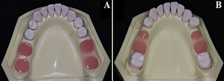
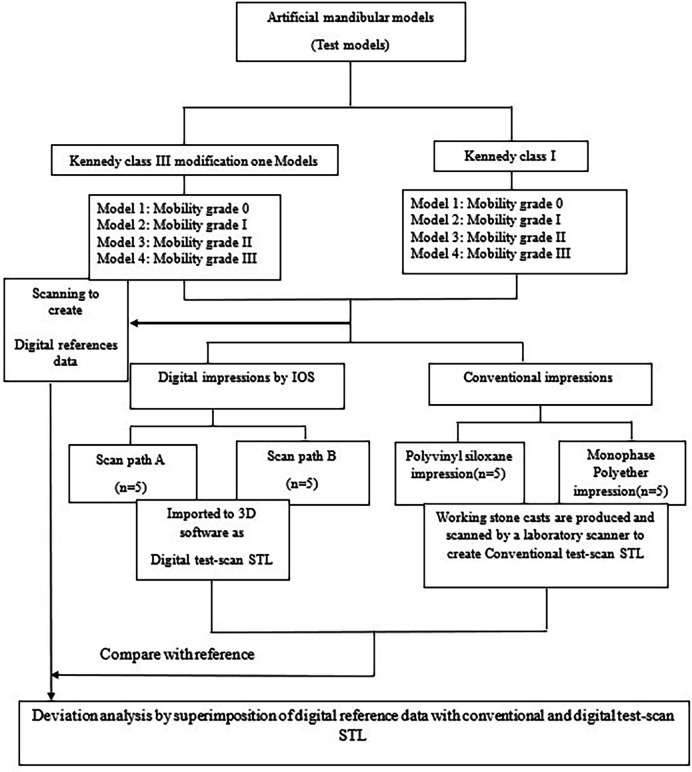
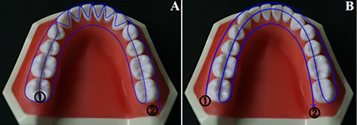
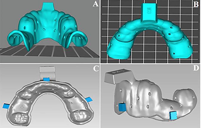
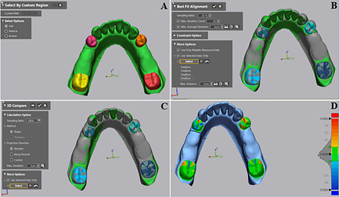
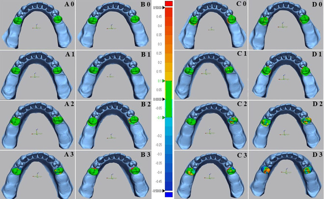
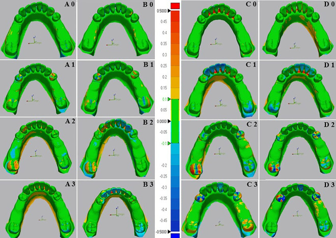
Eight lower Kennedy class I and class III models were employed as test models. The abutment teeth in these models were subjected to various mobility grades, according to the Miller classification. Reference data was generated by scanning the test models using an extra-oral laboratory scanner. An intra-oral scanner (TRIOS 4; V21; 3Shape A/S) was used to obtain ten digital impressions following two different scanning paths (Scan path A and Scan path B). For conventional impressions, two impression materials (Monophase polyether and Polyvinyl siloxane) were used to create ten impressions with a one-step technique. Working stone casts were produced and converted to digital data. Accuracy was assessed by analyzing the deviation between test data (digital and conventional data) and the reference data using 3D software (Geomagic Control X). The data was analysis using sequential tests, including two-way and one-way ANOVA, and paired t-tests (p < 0.05).
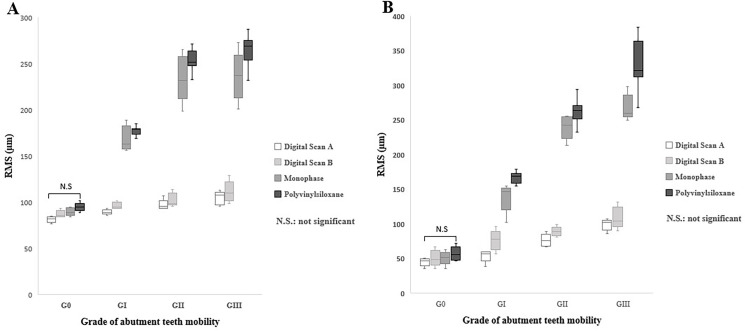
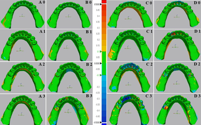
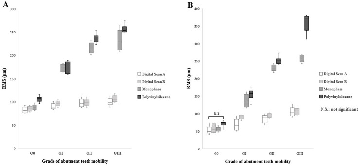
Digital impressions obtained through an intra-oral scanner exhibited significantly higher accuracy. Within the digital impression category, those recommended by the manufacturer obtained using scan path A showed lower deviations than those acquired through scan path B. Considering the degree of tooth mobility, models with GII and GIII mobile RPD abutment teeth displayed significantly higher deviations (p < 0.001) than those with G0, GI across all impression techniques. The accuracy of conventional impressions with GII and GIII mobility was clinically unacceptable (deviation >200µm).
For partially edentulous cases with mobile abutment teeth, digital impressions exhibited superior accuracy for G0, GI. Following the manufacturer-recommended scanning protocol in scan path A can improve the accuracy of impressions. Furthermore, if there is persistent mobility, particularly in GII and GIII, the use of final conventional impressions is forbidden.
牙周修复体或可摘局部义齿是治疗伴有牙周问题的部分牙列缺失的重要方法。本研究旨在评估在有活动基牙的部分无牙颌牙槽嵴中,通过口腔内扫描仪采用不同扫描路径获得的数字印模与传统印模的准确性。
采用八个下颌肯尼迪I类和III类模型作为测试模型。根据米勒分类法,这些模型中的基牙具有不同的松动度等级。通过使用口外实验室扫描仪扫描测试模型生成参考数据。使用口腔内扫描仪(TRIOS 4;V21;3Shape A/S)按照两种不同的扫描路径(扫描路径A和扫描路径B)获取十个数字印模。对于传统印模,使用两种印模材料(单相聚醚和聚乙烯基硅氧烷)采用一步法制作十个印模。制作工作石膏模型并转换为数字数据。使用3D软件(Geomagic Control X)通过分析测试数据(数字数据和传统数据)与参考数据之间的偏差来评估准确性。使用包括双向和单向方差分析以及配对t检验(p < 0.05)的序贯检验对数据进行分析。
通过口腔内扫描仪获得的数字印模显示出显著更高的准确性。在数字印模类别中,使用扫描路径A按照制造商推荐获得的印模显示出的偏差低于通过扫描路径B获得的印模。考虑到牙齿松动程度,在所有印模技术中,具有GII和GIII类活动可摘局部义齿基牙的模型显示出的偏差显著高于具有G0、GI类基牙的模型(p < 0.001)。具有GII和GIII类松动度的传统印模的准确性在临床上是不可接受的(偏差>200µm)。
对于有活动基牙的部分牙列缺失病例,数字印模在G0、GI类情况下显示出更高的准确性。按照扫描路径A中制造商推荐的扫描方案可以提高印模的准确性。此外,如果存在持续的松动,特别是在GII和GIII类情况下,禁止使用最终的传统印模。