Li Ye, Lv Xinna, Xu Xiaoyue, Zheng Ziwei, Lu Yiyan, Yao Zhijie, Hou Dailun
Department of Radiology, Shandong Provincial Hospital Affiliated to Shandong First Medical University, Jinan, China.
Department of Radiology, Beijing Chest Hospital, Capital Medical University, Beijing, China.
Quant Imaging Med Surg. 2025 Aug 1;15(8):6751-6762. doi: 10.21037/qims-2024-2671. Epub 2025 Jul 30.
The early prediction of treatment response for EGFR-tyrosine kinase inhibitors (EGFR-TKIs) is critical to guiding therapy in patients with metastatic non-small cell lung cancer (NSCLC). This study aimed to develop a magnetic resonance imaging (MRI)-based radiomics model based on intratumoral and peritumoral regions to assess the response of patients with metastatic NSCLC to EGFR-TKIs.
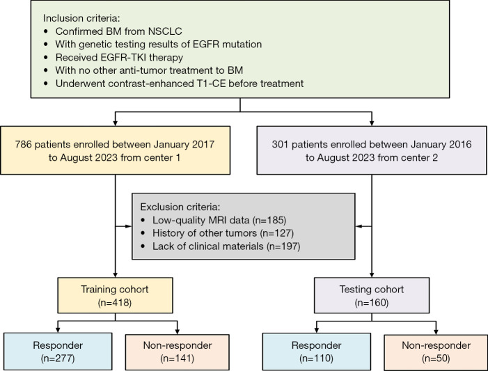
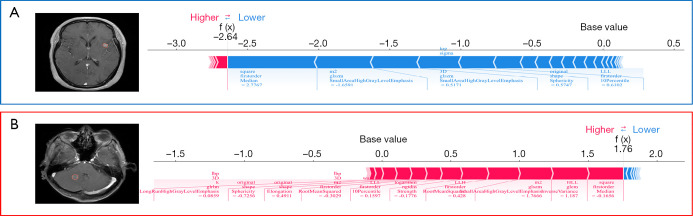
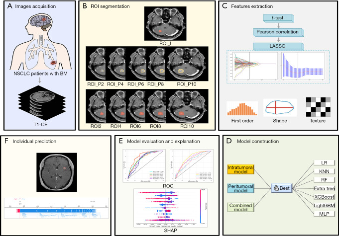
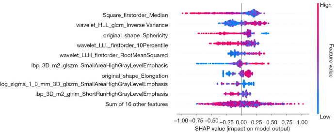
We retrospectively recruited 418 and 160 patients with brain metastases (BMs) from -mutant NSCLC who received EGFR-TKI therapy from hospital 1 and hospital 2, respectively. The intratumoral region of interest (ROI_I) was manually segmented for contrast-enhanced T1-weighted (T1-CE) imaging. Five peritumoral ROIs (ROI_P) at 2-, 4-, 6-, 8-, and 10-mm expansions along ROI_I were defined, and combined ROIs (ROI_I and ROI_P) were automatically generated. The least absolute shrinkage and selection operator (LASSO) was used to select the most predictive features, which was followed by the construction of radiomics models (the ROI_I model, ROI_P model, and the combined model). The area under the curve (AUC) and Shapley method were used to validate the performance of the models and explain the best models.
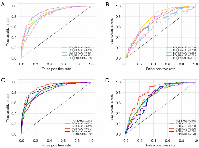
The combined intratumoral and peritumoral 6-mm regions achieved the best performance, with AUCs of 0.913 and 0.826 in the training and test cohort. The ROI_I model also demonstrated a degree of classification power in both the training and test cohort, with AUCs of 0.868 and 0.762, respectively.
As compared to models consisting of intratumoral or peritumoral radiomics features alone, the model combining intratumoral and peritumoral radiomics features achieved better performance in predicting therapeutic response to EGFR-TKIs. The optimal combined region model with 6-mm peritumoral expansion along the tumor may benefit the clinical treatment of NSCLC.
表皮生长因子受体-酪氨酸激酶抑制剂(EGFR-TKIs)治疗反应的早期预测对于指导转移性非小细胞肺癌(NSCLC)患者的治疗至关重要。本研究旨在基于肿瘤内和肿瘤周围区域开发一种基于磁共振成像(MRI)的放射组学模型,以评估转移性NSCLC患者对EGFR-TKIs的反应。
我们分别从医院1和医院2回顾性招募了418例和160例来自EGFR突变型NSCLC且接受EGFR-TKI治疗的脑转移(BM)患者。在对比增强T1加权(T1-CE)成像上手动分割肿瘤内感兴趣区域(ROI_I)。沿着ROI_I定义了5个在2、4、6、8和10毫米扩展的肿瘤周围ROI(ROI_P),并自动生成组合ROI(ROI_I和ROI_P)。使用最小绝对收缩和选择算子(LASSO)选择最具预测性的特征,随后构建放射组学模型(ROI_I模型、ROI_P模型和组合模型)。使用曲线下面积(AUC)和Shapley方法验证模型性能并解释最佳模型。
肿瘤内和肿瘤周围6毫米区域的组合表现最佳,在训练和测试队列中的AUC分别为0.913和0.826。ROI_I模型在训练和测试队列中也表现出一定程度的分类能力,AUC分别为0.868和0.762。
与仅由肿瘤内或肿瘤周围放射组学特征组成的模型相比,结合肿瘤内和肿瘤周围放射组学特征的模型在预测EGFR-TKIs治疗反应方面表现更好。沿肿瘤周围扩展6毫米的最佳组合区域模型可能有益于NSCLC的临床治疗。