• 文献检索
  • 文档翻译
  • 深度研究
  • 学术资讯
  • Suppr Zotero 插件Zotero 插件
  • 邀请有礼
  • 套餐&价格
  • 历史记录
应用&插件
Suppr Zotero 插件Zotero 插件浏览器插件Mac 客户端Windows 客户端微信小程序
定价
高级版会员购买积分包购买API积分包
服务
文献检索文档翻译深度研究API 文档MCP 服务
关于我们
关于 Suppr公司介绍联系我们用户协议隐私条款
关注我们

Suppr 超能文献

核心技术专利:CN118964589B侵权必究
粤ICP备2023148730 号-1Suppr @ 2026

文献检索

告别复杂PubMed语法,用中文像聊天一样搜索,搜遍4000万医学文献。AI智能推荐,让科研检索更轻松。

立即免费搜索

文件翻译

保留排版,准确专业,支持PDF/Word/PPT等文件格式,支持 12+语言互译。

免费翻译文档

深度研究

AI帮你快速写综述,25分钟生成高质量综述,智能提取关键信息,辅助科研写作。

立即免费体验

玻璃体视网膜界面异常行玻璃体切割术后视网膜内微囊肿的正面光学相干断层扫描及微视野分析

En-Face OCT and Microperimetric Analysis of Intraretinal Microcysts in Eyes After Pars Plana Vitrectomy for Epiretinal Membrane.

作者信息

Kaluzny Jakub J, Zabel Przemyslaw, Suwala Karolina Anna, Jaworski Damian, Gebska-Toloczko Martyna, Woznicki Krzysztof, Pek-Grzybowska Renata

机构信息

Department of Sensory Organ Studies, Nicolaus Copernicus University, Collegium Medicum, Bydgoszcz, Poland.

Oftalmika Eye Hospital, Bydgoszcz, Poland.

出版信息

Clin Ophthalmol. 2025 Aug 7;19:2587-2593. doi: 10.2147/OPTH.S524057. eCollection 2025.

DOI:10.2147/OPTH.S524057
PMID:40792187
原文链接:https://pmc.ncbi.nlm.nih.gov/articles/PMC12338324/
Abstract

PURPOSE

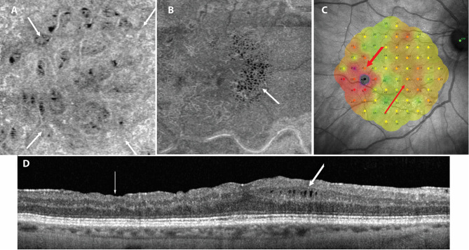
To compare the location of microcystic macular edema (MME) with areas of retinal nerve fiber layer (RNFL) damage in the macula detected on en-face SDOCT in eyes that underwent pars plana vitrectomy (PPV) due to the epiretinal membrane (ERM).

PATIENTS AND METHODS

Thirty-five eyes were enrolled at least 6 months after PPV with removal of ERM and inner limiting membrane (ILM). In each eye, en-face SDOCT and microperimetry were performed. The area of RNFL damage was measured and compared with the position of MME and correlated with the volume of retinal layers and retinal sensitivity (AT).

RESULTS

MME was observed in 17 eyes (48.6%) in the area devoid of ILM, often in places with arcuate damage to the RNFL bundle. The mean area of RNFL damage in eyes with MME was 9.03 ± 5.3 mm and was significantly larger than in eyes where microcysts were not present, where it measured 3.92 ± 3.3 mm. A significant negative correlation was observed between the area of RNFL damage and GCL volume and AT.

CONCLUSION

The topographic analysis of the MME position in eyes after PPV due to ERM confirmed the association of this pathology with ganglion cells and RNFL damage related to the removal of the ILM and ERM. There are probably two pathways leading to the development of MME: one starting from Muller cell damage during ILM peeling and the other due to retrograde death of ganglion cells in the areas of arcuate RNFL defects.

摘要

目的

比较因视网膜前膜(ERM)接受玻璃体切割术(PPV)的眼中,微囊性黄斑水肿(MME)的位置与经正面扫描光相干断层扫描(SDOCT)检测到的黄斑区视网膜神经纤维层(RNFL)损伤区域。

患者和方法

纳入35只在PPV联合ERM及内界膜(ILM)切除术后至少6个月的眼睛。对每只眼睛进行正面SDOCT和微视野检查。测量RNFL损伤面积,并与MME的位置进行比较,同时与视网膜各层体积和视网膜敏感度(AT)相关联。

结果

17只眼睛(48.6%)在无ILM的区域观察到MME,常出现在RNFL束呈弓形损伤的部位。存在MME的眼睛中RNFL损伤的平均面积为9.03±5.3平方毫米,显著大于无微囊肿的眼睛,后者为3.92±3.3平方毫米。观察到RNFL损伤面积与神经节细胞层(GCL)体积和AT之间存在显著负相关。

结论

对因ERM接受PPV术后眼睛中MME位置的地形图分析证实了这种病变与神经节细胞以及与ILM和ERM切除相关的RNFL损伤之间的关联。可能有两条导致MME发生的途径:一条始于ILM剥离过程中Müller细胞的损伤,另一条是由于弓形RNFL缺损区域神经节细胞的逆行性死亡。

https://cdn.ncbi.nlm.nih.gov/pmc/blobs/3862/12338324/0b469aa4c75c/OPTH-19-2587-g0002.jpg
https://cdn.ncbi.nlm.nih.gov/pmc/blobs/3862/12338324/8b974deb0d44/OPTH-19-2587-g0001.jpg
https://cdn.ncbi.nlm.nih.gov/pmc/blobs/3862/12338324/0b469aa4c75c/OPTH-19-2587-g0002.jpg
https://cdn.ncbi.nlm.nih.gov/pmc/blobs/3862/12338324/8b974deb0d44/OPTH-19-2587-g0001.jpg
https://cdn.ncbi.nlm.nih.gov/pmc/blobs/3862/12338324/0b469aa4c75c/OPTH-19-2587-g0002.jpg

相似文献

1
En-Face OCT and Microperimetric Analysis of Intraretinal Microcysts in Eyes After Pars Plana Vitrectomy for Epiretinal Membrane.玻璃体视网膜界面异常行玻璃体切割术后视网膜内微囊肿的正面光学相干断层扫描及微视野分析
Clin Ophthalmol. 2025 Aug 7;19:2587-2593. doi: 10.2147/OPTH.S524057. eCollection 2025.
2
Comparison of Structural Changes after Epiretinal Membrane Peeling between Nonglaucomatous Eyes and Eyes with Open-Angle Glaucoma.非青光眼性眼与开角型青光眼患者视网膜前膜剥脱术后结构变化的比较
Ophthalmol Glaucoma. 2025 Jul-Aug;8(4):351-359. doi: 10.1016/j.ogla.2025.03.001. Epub 2025 Mar 7.
3
Ganglion Cell Layer Thickness as a Biomarker for Amyotrophic Lateral Sclerosis Functional Outcome: An OCT study.神经节细胞层厚度作为肌萎缩侧索硬化症功能预后的生物标志物:一项光学相干断层扫描研究。
Rom J Ophthalmol. 2025 Apr-Jun;69(2):200-207. doi: 10.22336/rjo.2025.32.
4
En Face OCT Imaging of Epiretinal Membranes Complicated by Internal Limiting Membrane Tears.伴有内界膜撕裂的视网膜前膜的正面光学相干断层扫描成像
Ophthalmol Retina. 2025 Jun 2. doi: 10.1016/j.oret.2025.05.031.
5
Vitrectomy with internal limiting membrane (ILM) peeling versus vitrectomy with no peeling for idiopathic full-thickness macular hole (FTMH).玻璃体切除术联合内界膜(ILM)剥除术与单纯玻璃体切除术治疗特发性全层黄斑裂孔(FTMH)的对比
Cochrane Database Syst Rev. 2013 Jun 5(6):CD009306. doi: 10.1002/14651858.CD009306.pub2.
6
The impact of epiretinal membrane stage and postoperative treatment on visual and anatomical outcomes following vitrectomy in eyes with preexisting macular edema.视网膜前膜分期及术后治疗对已有黄斑水肿的眼睛玻璃体切除术后视觉和解剖学转归的影响。
Int J Retina Vitreous. 2025 Jul 1;11(1):74. doi: 10.1186/s40942-025-00697-y.
7
Long-term optical coherence tomography changes and visual outcomes after vitrectomy for epiretinal proliferation: lamellar holes and full-thickness macular holes.视网膜前增殖性病变玻璃体切除术后的长期光学相干断层扫描变化及视觉预后:板层孔和全层黄斑裂孔
Int J Retina Vitreous. 2025 Jul 24;11(1):83. doi: 10.1186/s40942-025-00711-3.
8
Idiopathic epiretinal membrane surgery with internal limiting membrane peeling: An optical coherence tomography angiography analysis of macular capillary plexus changes.特发性视网膜前膜手术联合内界膜剥除术:黄斑毛细血管丛变化的光学相干断层扫描血管造影分析
Eur J Ophthalmol. 2025 Jul;35(4):1394-1401. doi: 10.1177/11206721241304139. Epub 2024 Dec 19.
9
Long-Shaft Vitrectomy Probe for Vitreoretinal Diseases in Highly Myopic Eyes: A Randomized Controlled Trial.高度近视眼玻璃体视网膜疾病的长轴玻璃体切除探头:一项随机对照试验
Ophthalmol Sci. 2025 Jun 16;5(5):100824. doi: 10.1016/j.xops.2025.100824. eCollection 2025 Sep-Oct.
10
Internal Limiting Membrane Peeling for Grade C Proliferative Vitreoretinopathy: An International Multicenter Study.C级增生性玻璃体视网膜病变的内界膜剥除术:一项国际多中心研究
Ophthalmol Retina. 2025 Jun 17. doi: 10.1016/j.oret.2025.06.006.

本文引用的文献

1
Microcystoid Macular Edema in Epiretinal Membrane: Not a Retrograde Maculopathy.视网膜前膜中的微囊样黄斑水肿:并非逆行性黄斑病变。
Am J Ophthalmol. 2025 Apr;272:48-57. doi: 10.1016/j.ajo.2024.12.027. Epub 2025 Jan 3.
2
Assessment of ganglion cell complex thickness and its correlation with retinal sensitivity using microperimetry 6 months after epiretinal membrane surgery.视网膜前膜手术后6个月,使用微视野计评估神经节细胞复合体厚度及其与视网膜敏感度的相关性。
Int J Retina Vitreous. 2024 Aug 23;10(1):58. doi: 10.1186/s40942-024-00576-y.
3
Correlation between retinal sensitivity assessed by microperimetry and structural abnormalities on optical coherence tomography after successful epiretinal membrane surgery.
成功的视网膜前膜手术后,微视野检查评估的视网膜敏感度与光学相干断层扫描上的结构异常之间的相关性。
Int J Retina Vitreous. 2024 Feb 29;10(1):24. doi: 10.1186/s40942-024-00542-8.
4
CLINICAL FEATURES AND PROGNOSIS IN IDIOPATHIC EPIRETINAL MEMBRANES WITH DIFFERENT TYPES OF INTRARETINAL CYSTOID SPACES.特发性视网膜内囊样水肿型黄斑前膜的临床特征及预后。
Retina. 2022 Oct 1;42(10):1874-1882. doi: 10.1097/IAE.0000000000003537.
5
Non-vasogenic cystoid maculopathies.非血管源性囊样黄斑病变。
Prog Retin Eye Res. 2022 Nov;91:101092. doi: 10.1016/j.preteyeres.2022.101092. Epub 2022 Aug 1.
6
Clinical and Optical Coherence Tomography Analysis of Intraretinal Microcysts in Patients with Epiretinal Membrane.视网膜内微囊在视网膜前膜患者中的临床和光相干断层扫描分析。
Semin Ophthalmol. 2021 Nov 17;36(8):787-793. doi: 10.1080/08820538.2021.1906915. Epub 2021 Apr 1.
7
MACULAR SENSITIVITY IN THE AREA OF INTERNAL LIMITING MEMBRANE PEELING IN EYES AFTER PARS PLANA VITRECTOMY WITH THE TEMPORAL INVERTED INTERNAL LIMITING MEMBRANE FLAP TECHNIQUE FOR A FULL-THICKNESS MACULAR HOLE.内界膜撕除术后颞侧反转式全内界膜瓣技术治疗全层黄斑孔术后眼内界膜撕除区黄斑敏感度
Retina. 2021 Aug 1;41(8):1627-1634. doi: 10.1097/IAE.0000000000003096.
8
Risk of newly developing visual field defect and neurodegeneration after pars plana vitrectomy for idiopathic epiretinal membrane.特发性视网膜内界膜剥离术后新出现视野缺损和神经退行性变的风险。
Br J Ophthalmol. 2021 Dec;105(12):1683-1687. doi: 10.1136/bjophthalmol-2020-317478. Epub 2020 Oct 5.
9
Inner Nuclear Layer Microcyst Configuration, Distribution, and Visual Prognosis in Patients With Epiretinal Membrane After Vitrectomy and Membrane Peeling.内核层微囊结构、分布与玻璃体切除联合视网膜内界膜剥除术后眼内膜患者视力预后的关系
Sci Rep. 2019 Aug 9;9(1):11570. doi: 10.1038/s41598-019-48097-1.
10
PATIENTS WITH EPIRETINAL MEMBRANES DISPLAY RETROGRADE MACULOPATHY AFTER SURGICAL PEELING OF THE INTERNAL LIMITING MEMBRANE.内界膜切开术后,视网膜前膜患者出现逆行性黄斑病变。
Retina. 2019 Nov;39(11):2132-2140. doi: 10.1097/IAE.0000000000002266.