Cover Christopher, Reddy Sujatha, Vazquez Alberto, Fukuda Mitsuhiro, Poplawsky Alexander J
Department of Radiology, University of Pittsburgh, McGowan Institute for Regenerative Medicine Building, Pittsburgh, PA, United States.
Department of Bioengineering, University of Pittsburgh, Pittsburgh, PA, United States.
Imaging Neurosci (Camb). 2025 Jan 2;3. doi: 10.1162/imag_a_00406. eCollection 2025.
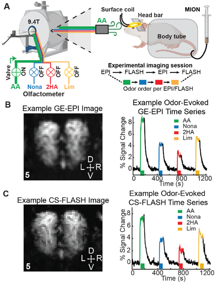
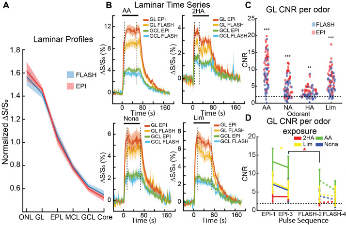
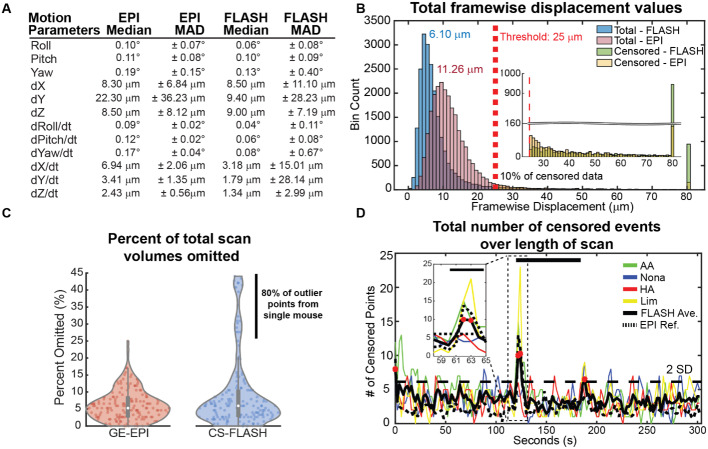
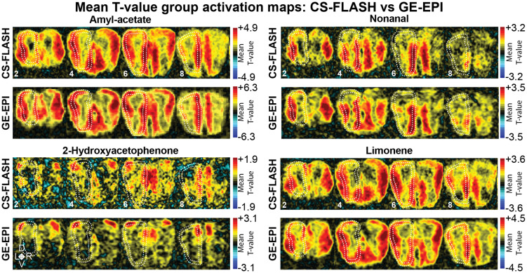
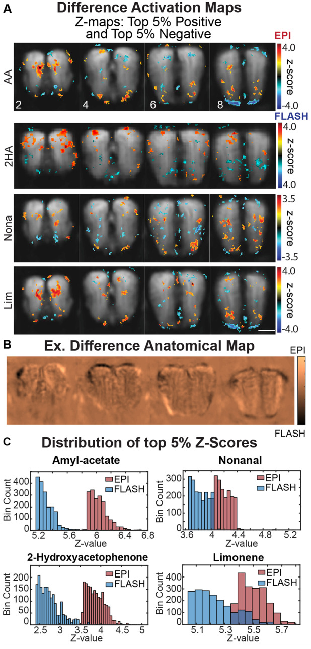
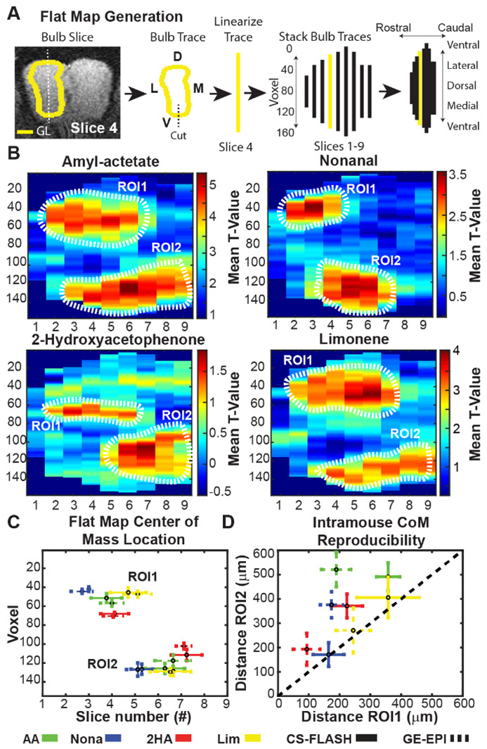
Awake rodent functional magnetic resonance imaging (fMRI) is increasingly becoming a reliable neuroimaging technique to study neuronal activity at both the whole-brain and high-resolution laminar scales. Prior studies have focused on developing acclimation protocols, experimental paradigms, and hardware to optimize outcomes. However, little effort has been made to address the impact of pulse sequence selection on detecting brain activation in awake fMRI experiments. In the current study, we compare gradient-echo echo planar imaging (GE-EPI) and compressed-sensing fast low-angle shot (CS-FLASH) sequences with cerebral blood volume-weighted (CBVw) contrast enhancement to investigate their sensitivity to hemodynamic activity in the olfactory bulb of awake rodents. Compared with GE-EPI, CS-FLASH had comparable motion parameters but was more sensitive to large motions, often resulting in corruption of the image quality. The use of framewise displacement as a motion censoring technique may over censor the data, requiring alternative approaches, such as spatial correlation censoring. CS-FLASH images were qualitatively sharper than GE-EPI; however, the contrast-to-noise ratio for odor activation was consistently greater for GE-EPI than for CS-FLASH that cannot be explained by olfactory adaptation alone. The activation maps of CS-FLASH to four different odors showed spatially unique patterns consistent with GE-EPI, but with lower z-scores or detection sensitivity. Activation maps were consistent with previously established histological findings. Additionally, odor-evoked laminar activation was greatest in the superficial layers that decreased with laminar depth, consistent with prior findings. We conclude that CS-FLASH produces sharper images with equivalent spatial activation maps to GE-EPI, albeit with lower statistical strength and contrast-to-noise ratio (CNR), and without being prohibited by motion-related image distortion.
清醒啮齿动物功能磁共振成像(fMRI)正日益成为一种可靠的神经成像技术,用于在全脑和高分辨率层状尺度上研究神经元活动。先前的研究主要集中在开发适应方案、实验范式和硬件以优化结果。然而,在清醒fMRI实验中,针对脉冲序列选择对检测脑激活的影响所做的工作很少。在本研究中,我们比较了具有脑血容量加权(CBVw)对比增强的梯度回波平面回波成像(GE-EPI)和压缩感知快速低角度激发(CS-FLASH)序列,以研究它们对清醒啮齿动物嗅球血流动力学活动的敏感性。与GE-EPI相比,CS-FLASH具有相当的运动参数,但对大运动更敏感,常常导致图像质量受损。使用逐帧位移作为运动检查技术可能会过度检查数据,需要采用替代方法,如空间相关性检查。CS-FLASH图像在定性上比GE-EPI更清晰;然而,GE-EPI的气味激活对比噪声比始终高于CS-FLASH,这不能仅用嗅觉适应来解释。CS-FLASH对四种不同气味的激活图显示出与GE-EPI一致的空间独特模式,但z分数或检测灵敏度较低。激活图与先前确立的组织学结果一致。此外,气味诱发的层状激活在表层最大,并随层深而降低,这与先前的发现一致。我们得出结论,CS-FLASH产生的图像比GE-EPI更清晰,具有等效的空间激活图,尽管统计强度和对比噪声比(CNR)较低,并且不受与运动相关的图像失真的影响。