• 文献检索
  • 文档翻译
  • 深度研究
  • 学术资讯
  • Suppr Zotero 插件Zotero 插件
  • 邀请有礼
  • 套餐&价格
  • 历史记录
应用&插件
Suppr Zotero 插件Zotero 插件浏览器插件Mac 客户端Windows 客户端微信小程序
定价
高级版会员购买积分包购买API积分包
服务
文献检索文档翻译深度研究API 文档MCP 服务
关于我们
关于 Suppr公司介绍联系我们用户协议隐私条款
关注我们

Suppr 超能文献

核心技术专利:CN118964589B侵权必究
粤ICP备2023148730 号-1Suppr @ 2026

文献检索

告别复杂PubMed语法,用中文像聊天一样搜索,搜遍4000万医学文献。AI智能推荐,让科研检索更轻松。

立即免费搜索

文件翻译

保留排版,准确专业,支持PDF/Word/PPT等文件格式,支持 12+语言互译。

免费翻译文档

深度研究

AI帮你快速写综述,25分钟生成高质量综述,智能提取关键信息,辅助科研写作。

立即免费体验

在深度学习分割的红外扫描激光眼底血管造影(IR-SLO)图像上使用可解释人工智能分析多发性硬化症患者的视网膜血管形态。

Analyzing Retinal Vessel Morphology in MS Using Interpretable AI on Deep Learning-Segmented IR-SLO Images.

作者信息

Soltanipour Asieh, Arian Roya, Aghababaei Ali, Ashtari Fereshteh, Zhou Yukun, Keane Pearse A, Kafieh Raheleh

机构信息

Medical Image and Signal Processing Research Center, Isfahan University of Medical Sciences, Isfahan 817467346, Iran.

Department of Engineering, Durham University, South Road, Durham DH1 3LE, UK.

出版信息

Bioengineering (Basel). 2025 Aug 6;12(8):847. doi: 10.3390/bioengineering12080847.

DOI:10.3390/bioengineering12080847
PMID:40868360
原文链接:https://pmc.ncbi.nlm.nih.gov/articles/PMC12384046/
Abstract

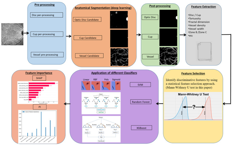
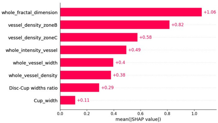
Multiple sclerosis (MS), a chronic disease of the central nervous system, is known to cause structural and vascular changes in the retina. Although optical coherence tomography (OCT) and fundus photography can detect retinal thinning and circulatory abnormalities, these findings are not specific to MS. This study explores the potential of Infrared Scanning-Laser-Ophthalmoscopy (IR-SLO) imaging to uncover vascular morphological features that may serve as MS-specific biomarkers. Using an age-matched, subject-wise stratified k-fold cross-validation approach, a deep learning model originally designed for color fundus images was adapted to segment optic disc, optic cup, and retinal vessels in IR-SLO images, achieving Dice coefficients of 91%, 94.5%, and 97%, respectively. This process included tailored pre- and post-processing steps to optimize segmentation accuracy. Subsequently, clinically relevant features were extracted. Statistical analyses followed by SHapley Additive exPlanations (SHAP) identified vessel fractal dimension, vessel density in zones B and C (circular regions extending 0.5-1 and 0.5-2 optic disc diameters from the optic disc margin, respectively), along with vessel intensity and width, as key differentiators between MS patients and healthy controls. These findings suggest that IR-SLO can non-invasively detect retinal vascular biomarkers that may serve as additional or alternative diagnostic markers for MS diagnosis, complementing current invasive procedures.

摘要

多发性硬化症(MS)是一种中枢神经系统的慢性疾病,已知会导致视网膜的结构和血管变化。尽管光学相干断层扫描(OCT)和眼底摄影可以检测到视网膜变薄和循环异常,但这些发现并非MS所特有。本研究探讨了红外扫描激光检眼镜(IR-SLO)成像揭示可能作为MS特异性生物标志物的血管形态特征的潜力。使用年龄匹配、按受试者分层的k折交叉验证方法,将最初为彩色眼底图像设计的深度学习模型进行调整,以分割IR-SLO图像中的视盘、视杯和视网膜血管,Dice系数分别达到91%、94.5%和97%。这一过程包括定制的预处理和后处理步骤,以优化分割精度。随后,提取临床相关特征。通过统计分析和SHapley加性解释(SHAP),确定血管分形维数、B区和C区(分别从视盘边缘延伸0.5-1和0.5-2个视盘直径的圆形区域)的血管密度,以及血管强度和宽度,是MS患者与健康对照之间的关键区分因素。这些发现表明,IR-SLO可以无创检测视网膜血管生物标志物,这些标志物可作为MS诊断的额外或替代诊断标志物,补充当前的侵入性检查方法。

https://cdn.ncbi.nlm.nih.gov/pmc/blobs/8338/12384046/e69c7775872f/bioengineering-12-00847-g010.jpg
https://cdn.ncbi.nlm.nih.gov/pmc/blobs/8338/12384046/ac17ce816db8/bioengineering-12-00847-g001.jpg
https://cdn.ncbi.nlm.nih.gov/pmc/blobs/8338/12384046/3d1c5e823b2e/bioengineering-12-00847-g002.jpg
https://cdn.ncbi.nlm.nih.gov/pmc/blobs/8338/12384046/b36458cac094/bioengineering-12-00847-g003.jpg
https://cdn.ncbi.nlm.nih.gov/pmc/blobs/8338/12384046/7ea75b1ab5cf/bioengineering-12-00847-g004.jpg
https://cdn.ncbi.nlm.nih.gov/pmc/blobs/8338/12384046/5b3744abad82/bioengineering-12-00847-g005.jpg
https://cdn.ncbi.nlm.nih.gov/pmc/blobs/8338/12384046/673ffd8940ae/bioengineering-12-00847-g006.jpg
https://cdn.ncbi.nlm.nih.gov/pmc/blobs/8338/12384046/6daa7409f43b/bioengineering-12-00847-g007.jpg
https://cdn.ncbi.nlm.nih.gov/pmc/blobs/8338/12384046/d15d6e474876/bioengineering-12-00847-g008.jpg
https://cdn.ncbi.nlm.nih.gov/pmc/blobs/8338/12384046/a2abf729d0fe/bioengineering-12-00847-g009.jpg
https://cdn.ncbi.nlm.nih.gov/pmc/blobs/8338/12384046/e69c7775872f/bioengineering-12-00847-g010.jpg

相似文献

1
Analyzing Retinal Vessel Morphology in MS Using Interpretable AI on Deep Learning-Segmented IR-SLO Images.在深度学习分割的红外扫描激光眼底血管造影(IR-SLO)图像上使用可解释人工智能分析多发性硬化症患者的视网膜血管形态。
Bioengineering (Basel). 2025 Aug 6;12(8):847. doi: 10.3390/bioengineering12080847.
2
SLO-Net: Enhancing Multiple Sclerosis Diagnosis Beyond Optical Coherence Tomography Using Infrared Reflectance Scanning Laser Ophthalmoscopy Images.SLO-Net:利用红外反射扫描激光检眼镜图像提升光学相干断层扫描技术之外的多发性硬化症诊断水平
Transl Vis Sci Technol. 2024 Jul 1;13(7):13. doi: 10.1167/tvst.13.7.13.
3
Ganglion Cell Layer Thickness as a Biomarker for Amyotrophic Lateral Sclerosis Functional Outcome: An OCT study.神经节细胞层厚度作为肌萎缩侧索硬化症功能预后的生物标志物:一项光学相干断层扫描研究。
Rom J Ophthalmol. 2025 Apr-Jun;69(2):200-207. doi: 10.22336/rjo.2025.32.
4
SLOctolyzer: Fully Automatic Analysis Toolkit for Segmentation and Feature Extracting in Scanning Laser Ophthalmoscopy Images.SLOctolyzer:用于扫描激光检眼镜图像分割和特征提取的全自动分析工具包。
Transl Vis Sci Technol. 2024 Nov 4;13(11):7. doi: 10.1167/tvst.13.11.7.
5
Artificial intelligence for diagnosing exudative age-related macular degeneration.人工智能在渗出性年龄相关性黄斑变性诊断中的应用。
Cochrane Database Syst Rev. 2024 Oct 17;10(10):CD015522. doi: 10.1002/14651858.CD015522.pub2.
6
Optic nerve head and fibre layer imaging for diagnosing glaucoma.用于诊断青光眼的视神经乳头和纤维层成像。
Cochrane Database Syst Rev. 2015 Nov 30;2015(11):CD008803. doi: 10.1002/14651858.CD008803.pub2.
7
Optical coherence tomography (OCT) for detection of macular oedema in patients with diabetic retinopathy.光学相干断层扫描(OCT)用于检测糖尿病视网膜病变患者的黄斑水肿。
Cochrane Database Syst Rev. 2011 Jul 6(7):CD008081. doi: 10.1002/14651858.CD008081.pub2.
8
Prescription of Controlled Substances: Benefits and Risks管制药品的处方:益处与风险
9
Optical coherence tomography (OCT) for detection of macular oedema in patients with diabetic retinopathy.光学相干断层扫描(OCT)用于检测糖尿病视网膜病变患者的黄斑水肿。
Cochrane Database Syst Rev. 2015 Jan 7;1(1):CD008081. doi: 10.1002/14651858.CD008081.pub3.
10
Optical coherence tomography and angiography reveal early retinal alterations in pediatric-onset multiple sclerosis.光学相干断层扫描和血管造影显示儿童期多发性硬化症患者早期视网膜改变。
Eur J Pediatr. 2025 Jun 6;184(7):398. doi: 10.1007/s00431-025-06234-1.

本文引用的文献

1
SLO-Net: Enhancing Multiple Sclerosis Diagnosis Beyond Optical Coherence Tomography Using Infrared Reflectance Scanning Laser Ophthalmoscopy Images.SLO-Net:利用红外反射扫描激光检眼镜图像提升光学相干断层扫描技术之外的多发性硬化症诊断水平
Transl Vis Sci Technol. 2024 Jul 1;13(7):13. doi: 10.1167/tvst.13.7.13.
2
Discrimination of multiple sclerosis using scanning laser ophthalmoscopy images with autoencoder-based feature extraction.基于自动编码器特征提取的扫描激光眼底镜图像对多发性硬化症的鉴别。
Mult Scler Relat Disord. 2024 Aug;88:105743. doi: 10.1016/j.msard.2024.105743. Epub 2024 Jun 21.
3
Ocular biomarkers of cognitive decline based on deep-learning retinal vessel segmentation.
基于深度学习视网膜血管分割的认知能力下降的眼部生物标志物。
BMC Geriatr. 2024 Jan 6;24(1):28. doi: 10.1186/s12877-023-04593-8.
4
Ocular microvascular complications in diabetic retinopathy: insights from machine learning.糖尿病视网膜病变的眼部微血管并发症:来自机器学习的见解。
BMJ Open Diabetes Res Care. 2024 Jan 2;12(1):e003758. doi: 10.1136/bmjdrc-2023-003758.
5
Evaluating Retinal Disease Diagnosis with an Interpretable Lightweight CNN Model Resistant to Adversarial Attacks.使用抗对抗攻击的可解释轻量级卷积神经网络模型评估视网膜疾病诊断
J Imaging. 2023 Oct 11;9(10):219. doi: 10.3390/jimaging9100219.
6
A Convolutional Neural Network Using Multimodal Retinal Imaging for Differentiation of Mild Cognitive Impairment from Normal Cognition.一种利用多模态视网膜成像技术区分轻度认知障碍与正常认知的卷积神经网络。
Ophthalmol Sci. 2023 Jun 25;4(1):100355. doi: 10.1016/j.xops.2023.100355. eCollection 2024 Jan-Feb.
7
Explainable artificial intelligence toward usable and trustworthy computer-aided diagnosis of multiple sclerosis from Optical Coherence Tomography.可解释人工智能在光学相干断层扫描中对多发性硬化症的计算机辅助诊断的应用与可信度。
PLoS One. 2023 Aug 7;18(8):e0289495. doi: 10.1371/journal.pone.0289495. eCollection 2023.
8
Discrimination of multiple sclerosis using OCT images from two different centers.使用来自两个不同中心的 OCT 图像对多发性硬化症进行鉴别。
Mult Scler Relat Disord. 2023 Sep;77:104846. doi: 10.1016/j.msard.2023.104846. Epub 2023 Jun 24.
9
Explainable AI for Retinoblastoma Diagnosis: Interpreting Deep Learning Models with LIME and SHAP.用于视网膜母细胞瘤诊断的可解释人工智能:使用LIME和SHAP解释深度学习模型
Diagnostics (Basel). 2023 Jun 1;13(11):1932. doi: 10.3390/diagnostics13111932.
10
Combining transfer learning with retinal lesion features for accurate detection of diabetic retinopathy.将迁移学习与视网膜病变特征相结合以实现糖尿病视网膜病变的准确检测。
Front Med (Lausanne). 2022 Nov 8;9:1050436. doi: 10.3389/fmed.2022.1050436. eCollection 2022.