Bogovska-Gigova Ralitsa, Ishkitiev Nikolay, Miteva Marina, Hristov Krasimir
Department of Pediatric Dentistry, Faculty of Dental Medicine, Medical University of Sofia, 1431 Sofia, Bulgaria.
Department of Chemistry and Biochemistry, Medical Faculty, Medical University of Sofia, 1431 Sofia, Bulgaria.
Materials (Basel). 2025 Aug 18;18(16):3863. doi: 10.3390/ma18163863.
This study aimed to evaluate the cytotoxicity of bulk-fill composite materials compared to conventional compomers on stem cells from human exfoliated deciduous teeth.
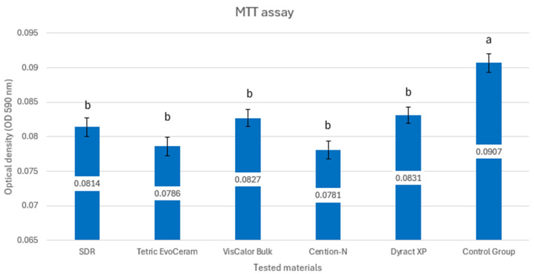
90 standardized resin composite discs (4 mm thick, 4 mm diameter) were fabricated using a 3D-printed plate, comprising four bulk-fill composites (SDR, Tetric EvoCeram Bulk-Fill, VisCalor Bulk, Cention-N) and one compomer (Dyract XP). Samples were polymerized per the manufacturer's instructions and sterilized. Stem cells were isolated from the pulp of exfoliated primary teeth. Cells were cultured and exposed to extracts of the composite materials soaked in culture medium for 24 h. Cytotoxicity was assessed using the MTT colorimetric assay, measuring cell viability via mitochondrial activity, and the Annexin V assay, quantifying apoptosis and necrosis via flow cytometry. Statistical analysis was performed using ANOVA and Tukey post hoc tests.
All materials significantly reduced cell viability compared to the control ( < 0.05), with optical density values indicating high cytotoxicity. Tetric EvoCeram exhibited the lowest necrosis and apoptosis levels, while Dyract XP showed the highest necrosis. Statistical analysis revealed no significant cytotoxicity differences among most bulk-fill composites ( < 0.05).
Bulk-fill composites and conventional compomer tested exhibit comparable and significant cytotoxic effects on stem cells from human exfoliated primary teeth pulp. While these materials offer clinical advantages in pediatric dentistry due to ease and speed of application, their use underscores the dilemma of balancing operative efficiency with biological safety, and their cytotoxic profiles should be taken into consideration prior to application.
本研究旨在评估大块充填复合树脂材料与人乳牙脱落后干细胞中传统复合体相比的细胞毒性。
使用3D打印板制作90个标准化树脂复合盘(4毫米厚,4毫米直径),其中包括四种大块充填复合树脂(SDR、Tetric EvoCeram Bulk-Fill、VisCalor Bulk、Cention-N)和一种复合体(Dyract XP)。样品按照制造商的说明进行聚合和灭菌。从脱落乳牙的牙髓中分离干细胞。将细胞培养并暴露于浸泡在培养基中的复合材料提取物中24小时。使用MTT比色法评估细胞毒性,通过线粒体活性测量细胞活力,使用Annexin V检测法通过流式细胞术定量凋亡和坏死。使用方差分析和Tukey事后检验进行统计分析。
与对照组相比,所有材料均显著降低了细胞活力(P < 0.05),光密度值表明细胞毒性高。Tetric EvoCeram表现出最低的坏死和凋亡水平,而Dyract XP表现出最高的坏死水平。统计分析显示,大多数大块充填复合树脂之间没有显著的细胞毒性差异(P < 0.05)。
所测试的大块充填复合树脂和传统复合体对人乳牙脱落后牙髓干细胞具有相当且显著的细胞毒性作用。虽然这些材料由于应用简便和速度快而在儿童牙科中具有临床优势,但它们的使用凸显了在手术效率与生物安全性之间取得平衡的困境,在应用前应考虑它们的细胞毒性特征。