Chattoraj D K, Inman R B
Proc Natl Acad Sci U S A. 1974 Feb;71(2):311-4. doi: 10.1073/pnas.71.2.311.
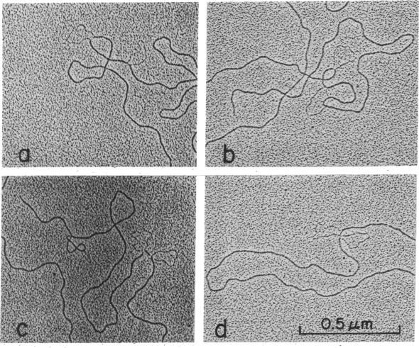
The physical position of vir37, a new immunity-insensitive mutant of Escherichia coli bacteriophage P2, was mapped by the electron microscopic heteroduplex method. In P2 vir37, a segment equivalent to 2.8% of P2 DNA is added. The addition was characterized as a tandem duplication of the segment occurring between 77.2 and 80.0% from the left end of P2 DNA (the right half of P2 DNA is arbitrarily defined, from denaturation map studies, as the half richer in A + T). The point of addition of the duplicated segment (the "novel-joint") was, thus, 80.0% from the left end of P2 DNA. On the basis of previous studies on P2 vir22, it was tentatively concluded that the physical and genetic maps of P2 are colinear. This conclusion is now further supported by physical and genetic data on P2 vir37.
通过电子显微镜异源双链法对大肠杆菌噬菌体P2的一种新的免疫不敏感突变体vir37的物理位置进行了定位。在P2 vir37中,添加了一段相当于P2 DNA 2.8%的片段。该添加片段的特征是在P2 DNA左端77.2%至80.0%之间发生的该片段的串联重复(根据变性图谱研究,P2 DNA的右半部分被任意定义为A + T含量较高的一半)。因此,重复片段的添加点(“新连接点”)位于P2 DNA左端的80.0%处。基于先前对P2 vir22的研究,初步得出结论,P2的物理图谱和遗传图谱是共线的。P2 vir37的物理和遗传数据现在进一步支持了这一结论。