• 文献检索
  • 文档翻译
  • 深度研究
  • 学术资讯
  • Suppr Zotero 插件Zotero 插件
  • 邀请有礼
  • 套餐&价格
  • 历史记录
应用&插件
Suppr Zotero 插件Zotero 插件浏览器插件Mac 客户端Windows 客户端微信小程序
定价
高级版会员购买积分包购买API积分包
服务
文献检索文档翻译深度研究API 文档MCP 服务
关于我们
关于 Suppr公司介绍联系我们用户协议隐私条款
关注我们

Suppr 超能文献

核心技术专利:CN118964589B侵权必究
粤ICP备2023148730 号-1Suppr @ 2026

文献检索

告别复杂PubMed语法,用中文像聊天一样搜索,搜遍4000万医学文献。AI智能推荐,让科研检索更轻松。

立即免费搜索

文件翻译

保留排版,准确专业,支持PDF/Word/PPT等文件格式,支持 12+语言互译。

免费翻译文档

深度研究

AI帮你快速写综述,25分钟生成高质量综述,智能提取关键信息,辅助科研写作。

立即免费体验

溶酶体与淀粉样蛋白早期沉积物密切关系的分析。实验性小鼠淀粉样变性的超微结构证据。

An analysis of the close relationship of lysosomes to early deposits of amyloid. Ultrastructural evidence in experimental mouse amyloidosis.

作者信息

Shirahama T, Cohen A S

出版信息

Am J Pathol. 1973 Oct;73(1):97-114.

PMID:4749216
原文链接:https://pmc.ncbi.nlm.nih.gov/articles/PMC1904042/
Abstract

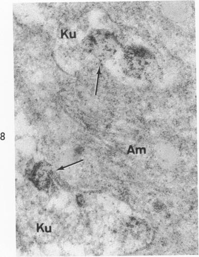
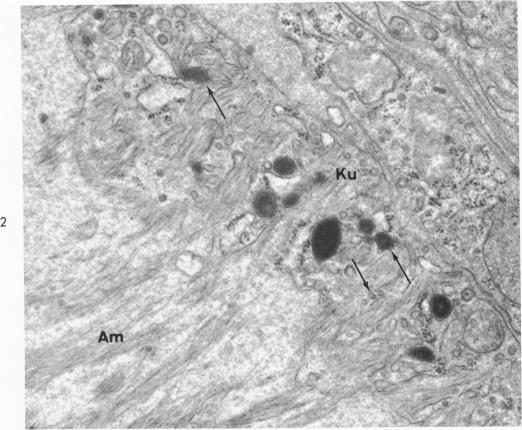
On the basis of recent morpholgic and biochemical studies which suggested the possible involvement of reticuloendothelial (RE) cells and proteolytic enzymes in amyloidogenesis, the present study was undertaken to examine the ultrastructural interrelationship between lysosomes and amyloid fibrils at the sites of very early amyloid deposition. In the spleen, liver and kidney of the experimental mouse model, foci of amyloid deposits were closely associated with the RE cells. The lysosomal enzyme activity, as marked by cytochemical demonstration of acid glycerophosphatase activity, was localized in the primary type lysosomes (as defined by their electron microscopic appearance), in the Golgi complexes, in the small cytoplasmic vesicles and occasionally widespread in the cytoplasm. They showed an intimate relationship to the amyloid fibrils. The findings were interpreted as favoring the hypothesis that the hydrolases play a role in amyloid fibril formation. The enzyme activity was also demonstrated in the secondary type lysosomes which occasionally contained amyloid fibrils that appeared to be phagocytized.

摘要

基于近期的形态学和生物化学研究表明网状内皮(RE)细胞和蛋白水解酶可能参与淀粉样变的发生,本研究旨在检查在淀粉样变非常早期沉积部位溶酶体与淀粉样纤维之间的超微结构相互关系。在实验小鼠模型的脾脏、肝脏和肾脏中,淀粉样沉积物灶与RE细胞密切相关。通过酸性甘油磷酸酶活性的细胞化学显示所标记的溶酶体酶活性,定位于初级类型的溶酶体(根据其电子显微镜外观定义)、高尔基体复合物、小细胞质囊泡中,偶尔广泛分布于细胞质中。它们与淀粉样纤维显示出密切关系。这些发现被解释为支持水解酶在淀粉样纤维形成中起作用的假说。在偶尔含有似乎被吞噬的淀粉样纤维的次级类型溶酶体中也证实了酶活性。

https://cdn.ncbi.nlm.nih.gov/pmc/blobs/4f78/1904042/ac1112f485dd/amjpathol00248-0115-b.jpg
https://cdn.ncbi.nlm.nih.gov/pmc/blobs/4f78/1904042/c7c9403107f2/amjpathol00248-0116-a.jpg
https://cdn.ncbi.nlm.nih.gov/pmc/blobs/4f78/1904042/9f2d144898f8/amjpathol00248-0116-b.jpg
https://cdn.ncbi.nlm.nih.gov/pmc/blobs/4f78/1904042/e6fb638a4699/amjpathol00248-0117-a.jpg
https://cdn.ncbi.nlm.nih.gov/pmc/blobs/4f78/1904042/65d5e8c4f2ee/amjpathol00248-0117-b.jpg
https://cdn.ncbi.nlm.nih.gov/pmc/blobs/4f78/1904042/3d9da4101d71/amjpathol00248-0117-c.jpg
https://cdn.ncbi.nlm.nih.gov/pmc/blobs/4f78/1904042/02d9b4459ff2/amjpathol00248-0113-a.jpg
https://cdn.ncbi.nlm.nih.gov/pmc/blobs/4f78/1904042/3ee86e72363b/amjpathol00248-0118-a.jpg
https://cdn.ncbi.nlm.nih.gov/pmc/blobs/4f78/1904042/efc25e41efa8/amjpathol00248-0114-a.jpg
https://cdn.ncbi.nlm.nih.gov/pmc/blobs/4f78/1904042/17ed6ace556c/amjpathol00248-0114-b.jpg
https://cdn.ncbi.nlm.nih.gov/pmc/blobs/4f78/1904042/32d3f14222de/amjpathol00248-0115-a.jpg
https://cdn.ncbi.nlm.nih.gov/pmc/blobs/4f78/1904042/ac1112f485dd/amjpathol00248-0115-b.jpg

相似文献

1
An analysis of the close relationship of lysosomes to early deposits of amyloid. Ultrastructural evidence in experimental mouse amyloidosis.溶酶体与淀粉样蛋白早期沉积物密切关系的分析。实验性小鼠淀粉样变性的超微结构证据。
Am J Pathol. 1973 Oct;73(1):97-114.
2
Intralysosomal formation of amyloid fibrils.淀粉样纤维在溶酶体内形成。
Am J Pathol. 1975 Oct;81(1):101-16.
3
Experimental amyloidosis using silver nitrate--electron microscopic study on the relationship between silver granules, amyloid fibrils and reticuloendothelial system.使用硝酸银的实验性淀粉样变性——银颗粒、淀粉样原纤维与网状内皮系统关系的电子显微镜研究
Acta Pathol Jpn. 1973 Aug;23(3):439-64. doi: 10.1111/j.1440-1827.1973.tb01217.x.
4
Experimental amyloidosis. Role of the hepatocytes and Kupffer cells in amyloid formation.实验性淀粉样变性。肝细胞和库普弗细胞在淀粉样蛋白形成中的作用。
Appl Pathol. 1985;3(1-2):78-87.
5
Experimental amyloidosis. II. Light and electron microscopic observations of liver.实验性淀粉样变性。II. 肝脏的光镜和电镜观察
Am J Pathol. 1964 Apr;44(4):629-44.
6
Pathological study on amyloidosis. Role of reticuloendothelial cells in inducing amyloidosis.淀粉样变性的病理学研究。网状内皮细胞在诱导淀粉样变性中的作用。
Acta Pathol Jpn. 1967 Feb;17(1):49-82.
7
Electron microscopy and electron microscopic autoradiography of splenic cell cultures from mice with amyloidosis.淀粉样变性小鼠脾细胞培养物的电子显微镜检查及电子显微镜放射自显影术
Lab Invest. 1969 Mar;20(3):234-42.
8
The SJL/J mouse: a new model for spontaneous age-associated amyloidosis. I. Morphologic and immunochemical aspects.SJL/J小鼠:自发性年龄相关性淀粉样变性的新模型。I. 形态学和免疫化学方面
Lab Invest. 1976 Jul;35(1):47-54.
9
Ultrastructural observations on the first amyloid to be found in the spleen of casein treated mice.对在酪蛋白处理小鼠脾脏中首次发现的淀粉样蛋白的超微结构观察。
Acta Pathol Microbiol Scand Suppl. 1972;233:141-50.
10
Deposition of amyloid in the liver of hamsters: an enzyme-histochemical and electron-microscopical study.仓鼠肝脏中淀粉样蛋白的沉积:酶组织化学与电子显微镜研究
Lab Anim. 1979 Jan;13(1):1-9. doi: 10.1258/002367779781071230.

引用本文的文献

1
AL(light chain)-amyloidogenesis by mesangial cells involves active participation of lysosomes: An ultrastructural study.系膜细胞的轻链淀粉样变形成涉及溶酶体的积极参与:一项超微结构研究。
Heliyon. 2023 Apr 7;9(4):e15190. doi: 10.1016/j.heliyon.2023.e15190. eCollection 2023 Apr.
2
Electron tomography reveals the fibril structure and lipid interactions in amyloid deposits.电子断层扫描揭示了淀粉样沉积物中的纤维结构和脂质相互作用。
Proc Natl Acad Sci U S A. 2016 May 17;113(20):5604-9. doi: 10.1073/pnas.1523496113. Epub 2016 May 2.
3
Basement membrane proteins and apolipoprotein E in growth hormone secreting adenomas and their correlation to amyloid: an immunoelectron microscopic study.

本文引用的文献

1
A SIMPLIFIED LEAD CITRATE STAIN FOR USE IN ELECTRON MICROSCOPY.一种用于电子显微镜的简化柠檬酸铅染色法。
J Cell Biol. 1965 May;25(2):407-8. doi: 10.1083/jcb.25.2.407.
2
LYTIC ACTIVITIES IN RENAL PROTEIN ABSORPTION DROPLETS. AN ELECTRON MICROSCOPICAL CYTOCHEMICAL STUDY.肾蛋白吸收液滴中的溶解活性。一项电子显微镜细胞化学研究。
J Cell Biol. 1964 Dec;23(3):519-52. doi: 10.1083/jcb.23.3.519.
3
GOLGI APPARATUS AND LYSOSOMES.高尔基体和溶酶体。
生长激素分泌性腺瘤中的基底膜蛋白和载脂蛋白E及其与淀粉样蛋白的相关性:一项免疫电子显微镜研究。
Endocr Pathol. 2000 Spring;11(1):49-56. doi: 10.1385/ep:11:1:49.
4
Redistribution of amyloid deposits.淀粉样沉积物的重新分布。
Am J Pathol. 1980 Jun;99(3):539-50.
5
Early glomerular lesions in amyloidosis. Electronmicroscopic findings.淀粉样变性中的早期肾小球病变。电子显微镜检查结果。
Virchows Arch A Pathol Anat Histol. 1981;390(3):259-72. doi: 10.1007/BF00496558.
6
Immunocytochemical identification of amyloid in formalin-fixed paraffin sections.福尔马林固定石蜡切片中淀粉样蛋白的免疫细胞化学鉴定
Histochemistry. 1981;72(2):161-71. doi: 10.1007/BF00517130.
7
Blockage of amyloid induction by colchicine in an animal model.秋水仙碱在动物模型中对淀粉样蛋白诱导的阻断作用。
J Exp Med. 1974 Oct 1;140(4):1102-7. doi: 10.1084/jem.140.4.1102.
8
Casein-induced experimental amyloidosis. III. Response to mitogens, allogeneic cells, and graft-versus-host reactions in the murine model.酪蛋白诱导的实验性淀粉样变性。III. 小鼠模型中对丝裂原、同种异体细胞及移植物抗宿主反应的应答
Immunology. 1974 Nov;27(5):953-63.
9
Islet amyloid in type 2 human diabetes mellitus and adult diabetic cats contains a novel putative polypeptide hormone.2型人类糖尿病和成年糖尿病猫的胰岛淀粉样蛋白含有一种新型的假定多肽激素。
Am J Pathol. 1987 Jun;127(3):414-7.
10
Amyloid neuropathy: immunocytochemical localization of intra- and extracellular immunoglobulin light chains.淀粉样变神经病:细胞内和细胞外免疫球蛋白轻链的免疫细胞化学定位
Acta Neuropathol. 1989;79(2):190-9. doi: 10.1007/BF00294378.
Fed Proc. 1964 Sep-Oct;23:1010-22.
4
PREPARATION OF THICK SECTIONS FOR CYTOCHEMISTRY AND ELECTRON MICROSCOPY BY A NON-FREEZING TECHNIQUE.用非冷冻技术制备用于细胞化学和电子显微镜检查的厚切片
Nature. 1963 Nov 16;200:691. doi: 10.1038/200691a0.
5
THE SITE OF FORMATION AND ULTRASTRUCTURE OF AMYLOID.淀粉样蛋白的形成部位与超微结构
Am J Pathol. 1963 Nov;43(5):837-54.
6
Improvements in epoxy resin embedding methods.环氧树脂包埋方法的改进。
J Biophys Biochem Cytol. 1961 Feb;9(2):409-14. doi: 10.1083/jcb.9.2.409.
7
Staining of tissue sections for electron microscopy with heavy metals.用重金属对组织切片进行电子显微镜染色。
J Biophys Biochem Cytol. 1958 Jul 25;4(4):475-8. doi: 10.1083/jcb.4.4.475.
8
The partial amino acid sequence of the major low molecular weight component of two human amyloid fibrils.两种人类淀粉样纤维主要低分子量成分的部分氨基酸序列。
FEBS Lett. 1972 Apr 15;22(1):121-123. doi: 10.1016/0014-5793(72)80235-7.
9
The major proteins of human and monkey amyloid substance: Common properties including unusual N-terminal amino acid sequences.人类和猴子淀粉样物质的主要蛋白质:共同特性,包括不寻常的N端氨基酸序列。
FEBS Lett. 1971 Dec 1;19(2):169-173. doi: 10.1016/0014-5793(71)80506-9.
10
The localization by electron microscopy of acid phosphatase activity in guinea pig macrophages.豚鼠巨噬细胞中酸性磷酸酶活性的电子显微镜定位
J Ultrastruct Res. 1966 Sep;16(1):96-108. doi: 10.1016/s0022-5320(66)80025-4.