• 文献检索
  • 文档翻译
  • 深度研究
  • 学术资讯
  • Suppr Zotero 插件Zotero 插件
  • 邀请有礼
  • 套餐&价格
  • 历史记录
应用&插件
Suppr Zotero 插件Zotero 插件浏览器插件Mac 客户端Windows 客户端微信小程序
定价
高级版会员购买积分包购买API积分包
服务
文献检索文档翻译深度研究API 文档MCP 服务
关于我们
关于 Suppr公司介绍联系我们用户协议隐私条款
关注我们

Suppr 超能文献

核心技术专利:CN118964589B侵权必究
粤ICP备2023148730 号-1Suppr @ 2026

文献检索

告别复杂PubMed语法,用中文像聊天一样搜索,搜遍4000万医学文献。AI智能推荐,让科研检索更轻松。

立即免费搜索

文件翻译

保留排版,准确专业,支持PDF/Word/PPT等文件格式,支持 12+语言互译。

免费翻译文档

深度研究

AI帮你快速写综述,25分钟生成高质量综述,智能提取关键信息,辅助科研写作。

立即免费体验

与衰老相关的胰腺形态变化。

Changes in pancreatic morphology associated with aging.

作者信息

Kreel L, Sandin B

出版信息

Gut. 1973 Dec;14(12):962-70. doi: 10.1136/gut.14.12.962.

DOI:10.1136/gut.14.12.962
PMID:4785285
原文链接:https://pmc.ncbi.nlm.nih.gov/articles/PMC1412863/
Abstract

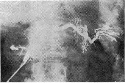
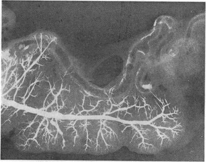
Retrograde pancreatography has been carried out at necropsy in 120 cases and the results have been analysed in statistical detail. With increasing age, changes in pancreatic anatomy occur which must not be taken to indicate pathology. These changes are: (1) low or ptotic position of the pancreas so that the papilla of Vater is below the level of L3; (2) calcification of the splenic and superior mesenteric arteries which produce calcific densities around the pancreas; (3) increasing width of main pancreatic duct along its whole length at about 8% per decade; in the elderly, widths of 1 cm can occur in the main duct in the head of the pancreas without evidence of obstruction; (4) formation of ductular ectasia which affects mainly the interlobular ductules but also intralobular ductules; (5) some ectatic ducts reach the dimensions of cysts, ie, 1-2 cm in diameter. OTHER MORPHOLOGICAL CHANGES WHICH HAVE BEEN DEMONSTRATED AND WHICH MAY PRODUCE DIFFICULTIES IN RADIOLOGICAL INTERPRETATION ARE: (a) narrowed ducts not due to stricture; (b) space-occupying lesions due to superior mesenteric artery, splenic artery, aorta, vertebral osteophytes, sympathetic ganglion, and lymph nodes; (c) metastases in the pancreas-these must be distinguished from primary pancreatic carcinoma. The implications of these findings for endoscopy and isotope pancreatic scanning will be mentioned.

摘要

已对120例尸体进行逆行胰管造影,并对结果进行了详细的统计学分析。随着年龄的增长,胰腺解剖结构会发生变化,但这些变化不一定意味着存在病理情况。这些变化包括:(1)胰腺位置低或下垂,致使Vater壶腹低于L3水平;(2)脾动脉和肠系膜上动脉钙化,在胰腺周围产生钙化密度影;(3)主胰管全长宽度每十年增加约8%;在老年人中,胰头主胰管宽度可达1cm,且无梗阻迹象;(4)形成导管扩张,主要影响小叶间导管,也影响小叶内导管;(5)一些扩张的导管达到囊肿大小,即直径1 - 2cm。其他已证实且可能在放射学解读中造成困难的形态学变化包括:(a)非狭窄性的导管狭窄;(b)由肠系膜上动脉、脾动脉、主动脉、椎体骨赘、交感神经节和淋巴结引起的占位性病变;(c)胰腺转移瘤——必须将其与原发性胰腺癌区分开来。将提及这些发现对内镜检查和同位素胰腺扫描的意义。

https://cdn.ncbi.nlm.nih.gov/pmc/blobs/e310/1412863/c7d942a671a4/gut00637-0059-a.jpg
https://cdn.ncbi.nlm.nih.gov/pmc/blobs/e310/1412863/a81b5d059354/gut00637-0055-a.jpg
https://cdn.ncbi.nlm.nih.gov/pmc/blobs/e310/1412863/2a9da72b776c/gut00637-0054-a.jpg
https://cdn.ncbi.nlm.nih.gov/pmc/blobs/e310/1412863/53c7febbe8a2/gut00637-0057-a.jpg
https://cdn.ncbi.nlm.nih.gov/pmc/blobs/e310/1412863/8bcc2b4ee8a3/gut00637-0057-b.jpg
https://cdn.ncbi.nlm.nih.gov/pmc/blobs/e310/1412863/061e7ec5d560/gut00637-0058-a.jpg
https://cdn.ncbi.nlm.nih.gov/pmc/blobs/e310/1412863/c7d942a671a4/gut00637-0059-a.jpg

相似文献

1
Changes in pancreatic morphology associated with aging.与衰老相关的胰腺形态变化。
Gut. 1973 Dec;14(12):962-70. doi: 10.1136/gut.14.12.962.
2
Pancreatic morphology--a combined radiological and pathological study.胰腺形态学——一项放射学与病理学的联合研究。
Clin Radiol. 1973 Apr;24(2):154-61. doi: 10.1016/s0009-9260(73)80074-1.
3
[Anatomy of the head of the pancreas and various limited resection procedures for intraductal papillary-mucinous tumors of the pancreas].[胰腺头部的解剖结构及胰腺导管内乳头状黏液性肿瘤的各种局限性切除手术]
Nihon Geka Gakkai Zasshi. 2003 Jun;104(6):460-70.
4
[Comparative radiological and morphological study of the human pancreas. III. Morphometric investigation of the major pancreatic duct (author's transl)].人体胰腺的比较放射学与形态学研究。III. 主胰管的形态计量学研究(作者译)
Z Gastroenterol. 1979 Apr;17(4):256-63.
5
[Intraorganic structure of blood vessels and ducts of the pancreas].
Khirurgiia (Mosk). 1989 Jul(7):72-5.
6
Endoscopic retrograde pancreatography: analysis of the normal pancreatogram.内镜逆行胰胆管造影术:正常胰管造影分析
Am J Dig Dis. 1976 Mar;21(3):263-9. doi: 10.1007/BF01095900.
7
Pancreatic duct diversity.
Pancreas. 2009 Apr;38(3):318-21. doi: 10.1097/MPA.0b013e31819081ba.
8
Problems in the diagnosis of pancreatic carcinoma by endoscopic retrograde cholangiopancreatography.内镜逆行胰胆管造影术诊断胰腺癌的问题
Gastrointest Radiol. 1977 Aug 18;2(1):61-5. doi: 10.1007/BF02256467.
9
Lymphatic system of the pancreas.胰腺的淋巴系统。
Microsc Res Tech. 1997;37(5-6):456-77. doi: 10.1002/(SICI)1097-0029(19970601)37:5/6<456::AID-JEMT9>3.0.CO;2-B.
10
Configurational anatomy of the pancreas: its surgical relevance from ontogenetic and comparative-anatomical viewpoints.胰腺的形态解剖学:从个体发生学和比较解剖学角度看其手术相关性。
J Hepatobiliary Pancreat Surg. 2003;10(1):48-56.

引用本文的文献

1
Exploring the variations of the pancreatic ductal system: a systematic review and meta-analysis of observational studies.探索胰腺导管系统的变异:观察性研究的系统评价和荟萃分析
Anat Cell Biol. 2024 Mar 31;57(1):31-44. doi: 10.5115/acb.23.148. Epub 2024 Feb 14.
2
Degenerative Changes in Aging Human Pelvic Lymph Nodes-A Reason to Rethink Staging and Therapy of Regional Malignancies?衰老人类盆腔淋巴结的退行性变化——是否是重新思考区域恶性肿瘤分期和治疗的原因?
Cancers (Basel). 2023 Sep 27;15(19):4754. doi: 10.3390/cancers15194754.
3
Pancreatic duct imaging during aging.

本文引用的文献

1
Calibre and appearance of the pancreatic ducts and relevant clinical problems. A roentgenographic and anatomical study.胰管的管径与外观及相关临床问题。一项X线造影与解剖学研究。
Acta Chir Scand. 1960 Feb 29;118:286-303.
2
Value of hypotonic duodenography as an adjunct to pancreatic scanning.低张十二指肠造影作为胰腺扫描辅助检查的价值。
Gut. 1969 Aug;10(8):665-73. doi: 10.1136/gut.10.8.665.
3
Pancreatic morphology--a combined radiological and pathological study.胰腺形态学——一项放射学与病理学的联合研究。
衰老过程中的胰管成像。
Endosc Ultrasound. 2023 Mar-Apr;12(2):200-212. doi: 10.4103/EUS-D-22-00119.
4
Causes of Exocrine Pancreatic Insufficiency Other Than Chronic Pancreatitis.除慢性胰腺炎外的外分泌性胰腺功能不全的病因
J Clin Med. 2021 Dec 10;10(24):5779. doi: 10.3390/jcm10245779.
5
The impact of age on the incidence and severity of post-endoscopic retrograde cholangiopancreatography pancreatitis.年龄对内镜逆行胰胆管造影术后胰腺炎发病率及严重程度的影响。
Ann Gastroenterol. 2018 Jan-Feb;31(1):96-101. doi: 10.20524/aog.2018.0203. Epub 2017 Oct 18.
6
Evolution of pancreas in aging: degenerative variation or early changes of disease?衰老过程中胰腺的演变:是退行性变化还是疾病的早期改变?
J Med Ultrason (2001). 2015 Apr;42(2):177-83. doi: 10.1007/s10396-014-0576-2. Epub 2014 Sep 13.
7
A study on the human pancreaticobiliary duct system and ampulla region with their clinical considerations.一项关于人类胰胆管系统及壶腹区域的研究及其临床考量。
Indian J Gastroenterol. 2015 Jan;34(1):58-62. doi: 10.1007/s12664-014-0526-3. Epub 2015 Mar 4.
8
Does an association exist between chronic pancreatitis and liver cirrhosis in alcoholic subjects?在酗酒者中,慢性胰腺炎和肝硬化之间存在关联吗?
World J Gastroenterol. 2008 Oct 28;14(40):6171-9. doi: 10.3748/wjg.14.6171.
9
Endoscopic ultrasonography.
J Gastrointest Surg. 2002 Mar-Apr;6(2):129-32. doi: 10.1016/s1091-255x(01)00056-7.
10
Is biliary lithiasis associated with pancreatographic changes?胆石症与胰管造影改变有关吗?
Gut. 1995 May;36(5):761-5. doi: 10.1136/gut.36.5.761.
Clin Radiol. 1973 Apr;24(2):154-61. doi: 10.1016/s0009-9260(73)80074-1.
4
The pancreas-radiographic demonstration of pancreatic morphology at autopsy.
Radiography. 1973 Jun;39(462):151-7.
5
Cannulation of the papilla of Vater by endoscopy and retrograde cholangiopancreatography (ERCP).通过内镜逆行胰胆管造影术(ERCP)对 Vater 乳头进行插管。
Gut. 1972 Dec;13(12):1014-25. doi: 10.1136/gut.13.12.1014.
6
The recognition and incidence of splenic artery aneurysms. A historical review.
Australas Radiol. 1972 Jun;16(2):126-36. doi: 10.1111/j.1440-1673.1972.tb01339.x.
7
The normal and abnormal pancreatic scan.
Q J Med. 1968 Oct;37(148):607-24.