• 文献检索
  • 文档翻译
  • 深度研究
  • 学术资讯
  • Suppr Zotero 插件Zotero 插件
  • 邀请有礼
  • 套餐&价格
  • 历史记录
应用&插件
Suppr Zotero 插件Zotero 插件浏览器插件Mac 客户端Windows 客户端微信小程序
定价
高级版会员购买积分包购买API积分包
服务
文献检索文档翻译深度研究API 文档MCP 服务
关于我们
关于 Suppr公司介绍联系我们用户协议隐私条款
关注我们

Suppr 超能文献

核心技术专利:CN118964589B侵权必究
粤ICP备2023148730 号-1Suppr @ 2026

文献检索

告别复杂PubMed语法,用中文像聊天一样搜索,搜遍4000万医学文献。AI智能推荐,让科研检索更轻松。

立即免费搜索

文件翻译

保留排版,准确专业,支持PDF/Word/PPT等文件格式,支持 12+语言互译。

免费翻译文档

深度研究

AI帮你快速写综述,25分钟生成高质量综述,智能提取关键信息,辅助科研写作。

立即免费体验

果胶梭菌孢子萌发的细胞学研究

Cytology of spore germination in Clostridium pectinovorum.

作者信息

Hoeniger J F, Headley C L

出版信息

J Bacteriol. 1968 Nov;96(5):1835-47. doi: 10.1128/jb.96.5.1835-1847.1968.

DOI:10.1128/jb.96.5.1835-1847.1968
PMID:4882027
原文链接:https://pmc.ncbi.nlm.nih.gov/articles/PMC315247/
Abstract

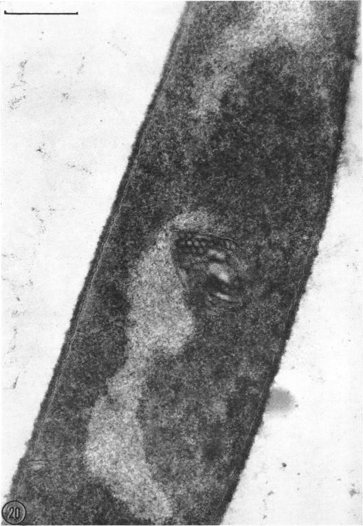
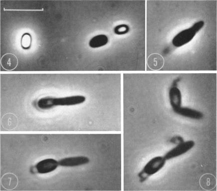
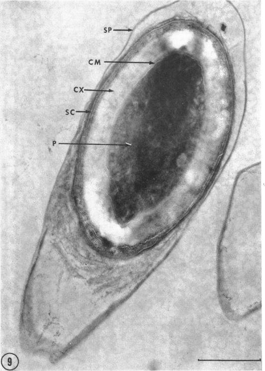
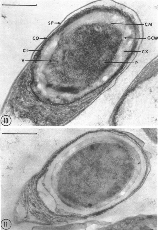
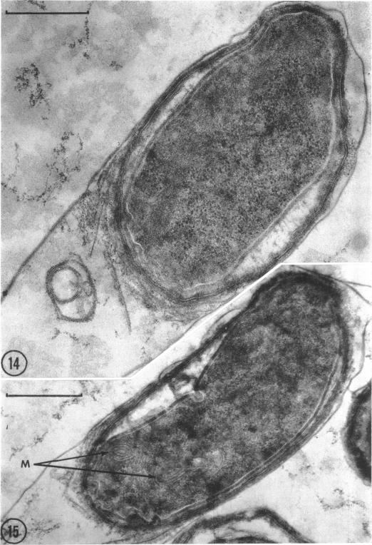
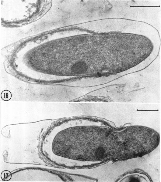
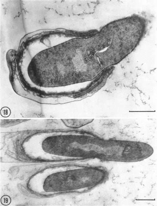
The process of spore germination in Clostridium pectinovorum has been followed by phase-contrast and electron microscopy. Unlike most other Bacillaceae, germination of this species takes place within the sporangium. Under phase-contrast, the spore darkens and swells slightly, and then the vegetative rod slips out through the end opposite the collar-like extension of the sporangium. In thin sections, a spore from an early stage in germination consists of a central protoplast, core membrane, germ cell wall, cortex, and two coats. Within a short period, the cortex disintegrates and the young cell develops. It possesses a large fibrillar nucleoplasm and several mesosomes. Subsequently, the young cell elongates, becomes somewhat deformed, and then emerges through a narrow aperture in the inflexible coats of the spore, finally rupturing the sporangium. Free vegetative cells of C. pectinovorum resemble in their structure other gram-positive rods.

摘要

通过相差显微镜和电子显微镜观察了果胶梭菌的孢子萌发过程。与大多数其他芽孢杆菌科不同,该菌种的萌发发生在孢子囊内。在相差显微镜下,孢子变暗并略有肿胀,然后营养杆从与孢子囊领状延伸相对的一端滑出。在薄片中,处于萌发早期的孢子由中央原生质体、核心膜、生殖细胞壁、皮层和两层外膜组成。在短时间内,皮层解体,幼细胞发育。它具有大的纤维状核质和几个中体。随后,幼细胞伸长,变得有些变形,然后通过孢子坚硬外膜上的一个窄孔出现,最终冲破孢子囊。果胶梭菌的游离营养细胞在结构上与其他革兰氏阳性杆菌相似。

https://cdn.ncbi.nlm.nih.gov/pmc/blobs/6770/315247/d0fed4522863/jbacter00589-0427-a.jpg
https://cdn.ncbi.nlm.nih.gov/pmc/blobs/6770/315247/8bc1c9b4a0e2/jbacter00589-0418-a.jpg
https://cdn.ncbi.nlm.nih.gov/pmc/blobs/6770/315247/049e1c78382b/jbacter00589-0419-a.jpg
https://cdn.ncbi.nlm.nih.gov/pmc/blobs/6770/315247/920954c04cc7/jbacter00589-0420-a.jpg
https://cdn.ncbi.nlm.nih.gov/pmc/blobs/6770/315247/39b744385235/jbacter00589-0422-a.jpg
https://cdn.ncbi.nlm.nih.gov/pmc/blobs/6770/315247/6935250aeb0c/jbacter00589-0423-a.jpg
https://cdn.ncbi.nlm.nih.gov/pmc/blobs/6770/315247/f969de3742b4/jbacter00589-0424-a.jpg
https://cdn.ncbi.nlm.nih.gov/pmc/blobs/6770/315247/6c9e192092a7/jbacter00589-0425-a.jpg
https://cdn.ncbi.nlm.nih.gov/pmc/blobs/6770/315247/764bd219f256/jbacter00589-0426-a.jpg
https://cdn.ncbi.nlm.nih.gov/pmc/blobs/6770/315247/d0fed4522863/jbacter00589-0427-a.jpg

相似文献

1
Cytology of spore germination in Clostridium pectinovorum.果胶梭菌孢子萌发的细胞学研究
J Bacteriol. 1968 Nov;96(5):1835-47. doi: 10.1128/jb.96.5.1835-1847.1968.
2
Ultrastructural changes associated with germination and outgrowth of an appendage-bearing clostridial spore.与带有附属物的梭菌芽孢萌发和生长相关的超微结构变化。
J Bacteriol. 1970 Mar;101(3):1038-45. doi: 10.1128/jb.101.3.1038-1045.1970.
3
Spore fine structure in Clostridium cochlearium.耳蜗梭菌的芽孢精细结构。
J Bacteriol. 1969 Nov;100(2):994-1001. doi: 10.1128/jb.100.2.994-1001.1969.
4
Ultrastructural aspects of spore germination and outgrowth in Clostridium sporogenes.产芽孢梭菌孢子萌发和生长的超微结构特征
Can J Microbiol. 1969 Sep;15(9):1061-5. doi: 10.1139/m69-189.
5
Apertures in the Clostridium sporogenes spore coat and exosporium align to facilitate emergence of the vegetative cell.生孢梭菌芽孢衣和芽孢外壁上的小孔对齐,以利于营养细胞的出现。
Food Microbiol. 2015 Oct;51:45-50. doi: 10.1016/j.fm.2015.04.013. Epub 2015 May 9.
6
Cytology of spore formation in Clostridium perfringens.产气荚膜梭菌孢子形成的细胞学研究
J Bacteriol. 1968 Nov;96(5):1818-34. doi: 10.1128/jb.96.5.1818-1834.1968.
7
Appendage development in Clostridium bifermentans.双酶梭菌的附属器发育
J Bacteriol. 1971 Apr;106(1):269-75. doi: 10.1128/jb.106.1.269-275.1971.
8
Bacterial spore appendages.细菌芽孢附属物。
CRC Crit Rev Microbiol. 1971 May;1(1):1-27. doi: 10.3109/10408417109104476.
9
[Structure of Clostridia of the butyric group. I. Sporulation of Clostridium butyricum and Clostridium saccharobutyricum].[丁酸梭菌属的结构。I. 丁酸梭菌和糖丁酸梭菌的孢子形成]
Ann Inst Pasteur (Paris). 1971 Jan;120(1):23-32.
10
MORPHOLOGY OF SPORE DEVELOPMENT IN CLOSTRIDIUM PECTINOVORUM.果胶分解梭菌孢子发育的形态学
J Bacteriol. 1962 Jul;84(1):104-14. doi: 10.1128/jb.84.1.104-114.1962.

引用本文的文献

1
Adaptive Strategies in a Poly-Extreme Environment: Differentiation of Vegetative Cells in and Resistance to Extreme Conditions.多极端环境中的适应性策略:营养细胞的分化及对极端条件的抗性
Front Microbiol. 2019 Feb 5;10:102. doi: 10.3389/fmicb.2019.00102. eCollection 2019.
2
Sporulation of Clostridium acetobutylicum P262 in a Defined Medium.丙酮丁醇梭菌 P262 在限定培养基中的孢子形成。
Appl Environ Microbiol. 1983 Apr;45(4):1389-93. doi: 10.1128/aem.45.4.1389-1393.1983.
3
Solvent Production and Morphological Changes in Clostridium acetobutylicum.

本文引用的文献

1
MORPHOLOGY OF SPORE DEVELOPMENT IN CLOSTRIDIUM PECTINOVORUM.果胶分解梭菌孢子发育的形态学
J Bacteriol. 1962 Jul;84(1):104-14. doi: 10.1128/jb.84.1.104-114.1962.
2
PRELIMINARY OBSERVATIONS ON THE GERMINATION OF THE ENDOSPORE IN BACILLUS MEGATHERIUM AND THE STRUCTURE OF THE SPORE COAT.巨大芽孢杆菌芽孢萌发及芽孢衣结构的初步观察
J Bacteriol. 1949 Jan;57(1):23-9. doi: 10.1128/jb.57.1.23-29.1949.
3
A Study, with the High-Voltage Electron Microscope, of the Endospore and Life Cycle of Bacillus mycoides.用高压电子显微镜对蕈状芽孢杆菌的芽孢和生命周期进行的研究。
溶剂生成和形态变化在丙酮丁醇梭菌中的研究。
Appl Environ Microbiol. 1982 Jun;43(6):1434-9. doi: 10.1128/aem.43.6.1434-1439.1982.
4
Ultrastructural changes associated with germination and outgrowth of an appendage-bearing clostridial spore.与带有附属物的梭菌芽孢萌发和生长相关的超微结构变化。
J Bacteriol. 1970 Mar;101(3):1038-45. doi: 10.1128/jb.101.3.1038-1045.1970.
5
Exosporium formation in sporulating cells of Clostridium botulinum 78A.肉毒梭菌78A芽孢形成细胞中的芽孢外壁形成
J Bacteriol. 1972 Oct;112(1):618-21. doi: 10.1128/jb.112.1.618-621.1972.
6
Fixation of mature spores of Clostridium botulinum.肉毒梭菌成熟孢子的固定
J Bacteriol. 1972 Mar;109(3):1295-7. doi: 10.1128/jb.109.3.1295-1297.1972.
7
Germination of spores from Clostridium botulinum B-aphis and Ba410.肉毒梭菌B型蚜虫株和Ba410株孢子的萌发
Appl Environ Microbiol. 1985 Oct;50(4):795-800. doi: 10.1128/aem.50.4.795-800.1985.
8
Ultrastructural changes during encystment and germination of Bdellovibrio sp.蛭弧菌在包囊形成和萌发过程中的超微结构变化
J Bacteriol. 1977 Jul;131(1):323-30. doi: 10.1128/jb.131.1.323-330.1977.
J Bacteriol. 1947 May;53(5):525-37. doi: 10.1128/jb.53.5.525-537.1947.
4
The use of lead citrate at high pH as an electron-opaque stain in electron microscopy.在电子显微镜检查中,将高pH值的柠檬酸铅用作电子不透明染色剂。
J Cell Biol. 1963 Apr;17(1):208-12. doi: 10.1083/jcb.17.1.208.
5
Electron microscope studies on ultrathin sections of spores of the Clostridium group, with special reference to the sporulation and germination process.关于梭菌属孢子超薄切片的电子显微镜研究,特别涉及孢子形成和萌发过程。
J Bacteriol. 1960 Jul;80(1):37-46. doi: 10.1128/jb.80.1.37-46.1960.
6
Electron microscopic studies on ultrathin sections of spores of Clostridium tetani and Clostridium histolyticum, with special reference to sporulation and spore germination process.破伤风梭菌和溶组织梭菌孢子超薄切片的电子显微镜研究,特别涉及孢子形成和孢子萌发过程。
Jpn J Microbiol. 1960 Apr;4:137-55. doi: 10.1111/j.1348-0421.1960.tb00162.x.
7
Study of germinating Bacillus cereus spores employing television microscopy of living cells and electron microscopy of ultrathin sections.利用活细胞电视显微镜和超薄切片电子显微镜对蜡样芽孢杆菌芽孢萌发的研究。
J Bacteriol. 1957 Aug;74(2):126-32. doi: 10.1128/jb.74.2.126-132.1957.
8
Nuclear division as observed in live bacteria by a new technique.通过一项新技术在活细菌中观察到的核分裂。
J Bacteriol. 1956 Apr;71(4):474-9. doi: 10.1128/jb.71.4.474-479.1956.
9
Biochemical changes occurring during the germination of bacterial spores.细菌芽孢萌发过程中发生的生化变化。
Biochem J. 1953 May;54(2):205-9. doi: 10.1042/bj0540205.
10
[Sporulation and germination in Clostridium butyricum M 55. Electron microscopic studies].[丁酸梭菌M 55中的孢子形成与萌发。电子显微镜研究]
Zentralbl Bakteriol Orig. 1966 Nov;201(3):373-95.