Murphy F A, Webb P A, Johnson K M, Whitfield S G, Chappell W A
J Virol. 1970 Oct;6(4):507-18. doi: 10.1128/JVI.6.4.507-518.1970.
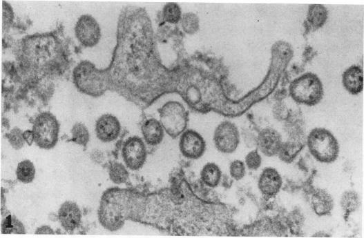
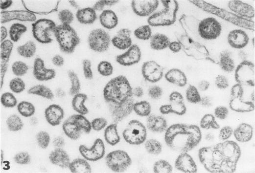
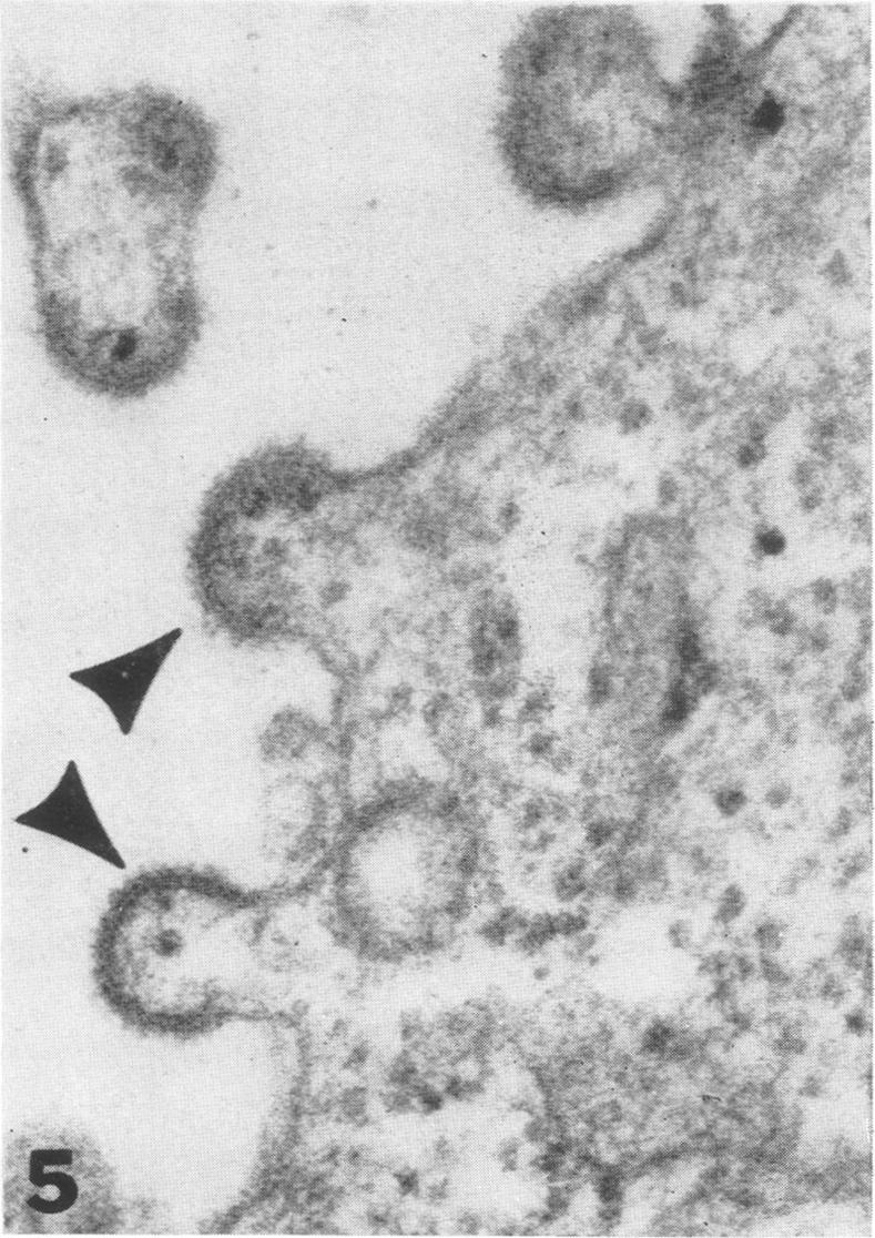
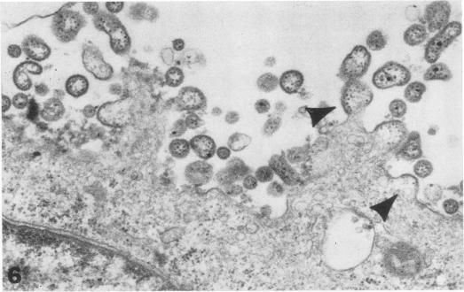
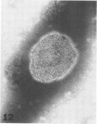
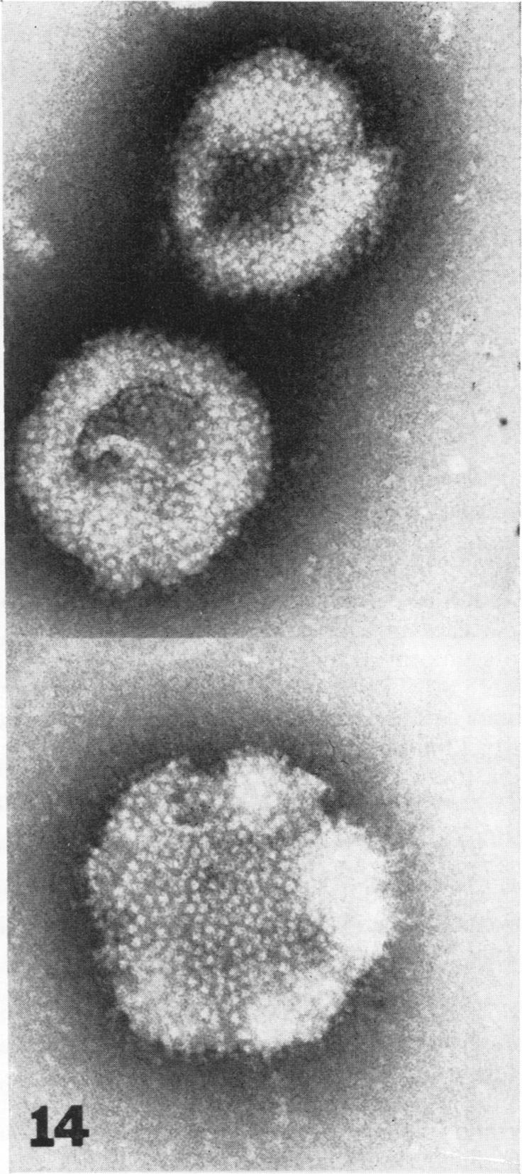
Thin-section electron microscopy was carried out on Vero green monkey kidney cell cultures infected with some viruses of the newly constituted arenovirus group. Junin, Machupo, Amapari, Pichinde, Parana, Tamiami, and Latino viruses were morphologically identical and indistinguishable from lymphocytic choriomeningitis virus, the prototype virus of the group. Virus particles were round, oval, or pleomorphic, 60 to 280 nm in diameter, and matured via budding from plasma membranes. Most characteristically, particles contained various amounts of homogeneous, 20- to 25-nm, dense granules; these granules in large masses also formed distinctive intracytoplasmic inclusions. In negative-contrast preparations from infected Vero cell culture supernatant fluids, several of the viruses appeared as pleomorphic membrane-bound forms with rather pronounced surface projections. Most particles were between 90 and 220 nm in diameter, although some reached 350 nm in their longest dimension. Internal structure was not resolved by negative-contrast electron microscopy. All observations supported the current delineation of a distinct arenovirus group.
对感染了新组建的沙粒病毒科某些病毒的非洲绿猴肾细胞培养物进行了超薄切片电子显微镜检查。胡宁病毒、马丘波病毒、阿马帕里病毒、皮钦德病毒、巴拉那病毒、塔米亚米病毒和拉蒂诺病毒在形态上相同,与该病毒科的原型病毒淋巴细胞性脉络丛脑膜炎病毒无法区分。病毒粒子呈圆形、椭圆形或多形性,直径为60至280纳米,通过从质膜出芽成熟。最典型的是,粒子含有不同数量的均匀的、直径为20至25纳米的致密颗粒;这些大量的颗粒还形成了独特的胞质内包涵体。在感染的非洲绿猴肾细胞培养物上清液的负染色制剂中,几种病毒呈现为多形性膜结合形式,表面突起相当明显。大多数粒子直径在90至220纳米之间,尽管有些粒子最长尺寸达到350纳米。负染色电子显微镜未分辨出内部结构。所有观察结果都支持目前对一个独特沙粒病毒科的划分。