Stackpole C W
J Virol. 1969 Jul;4(1):75-93. doi: 10.1128/JVI.4.1.75-93.1969.
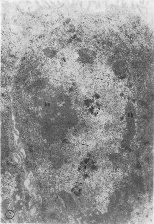
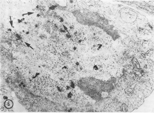
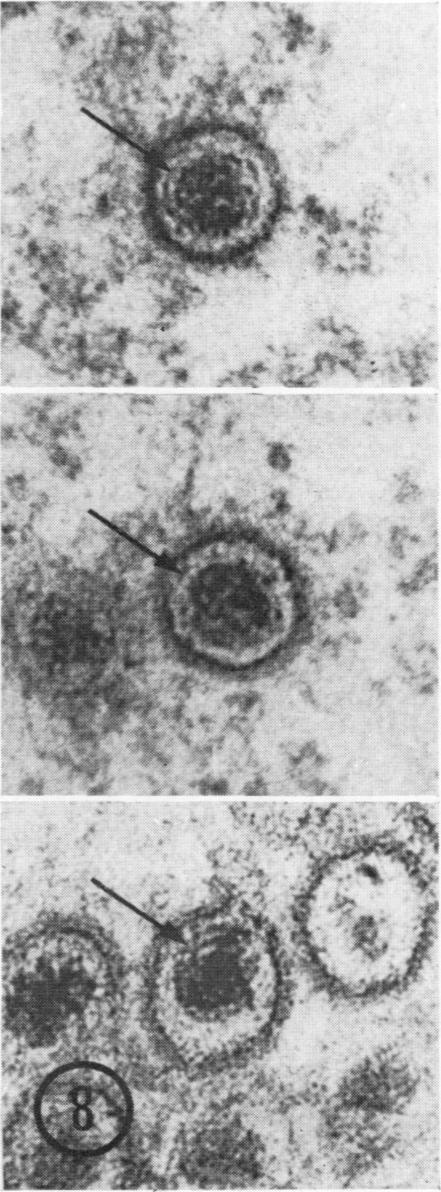
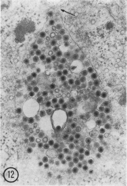
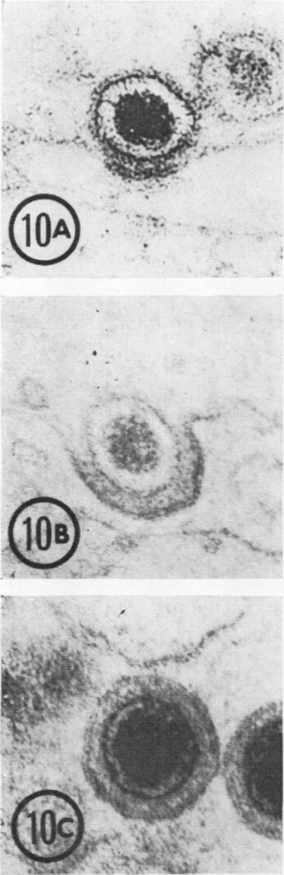
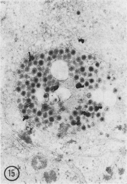
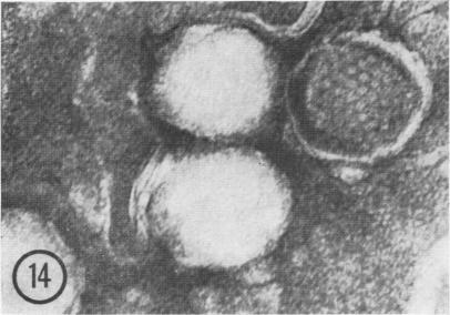
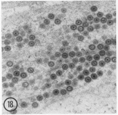
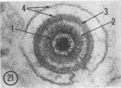
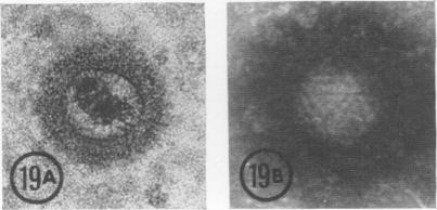
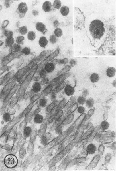
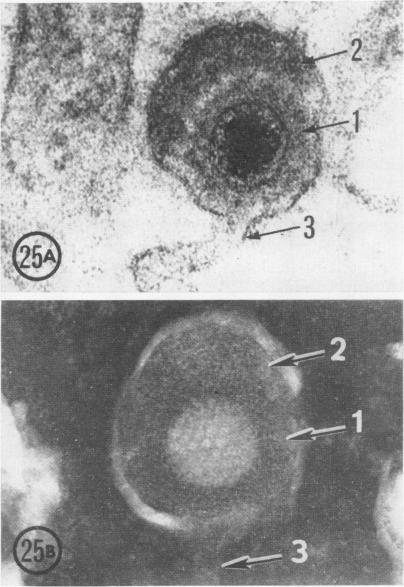
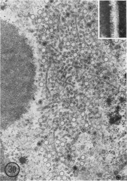
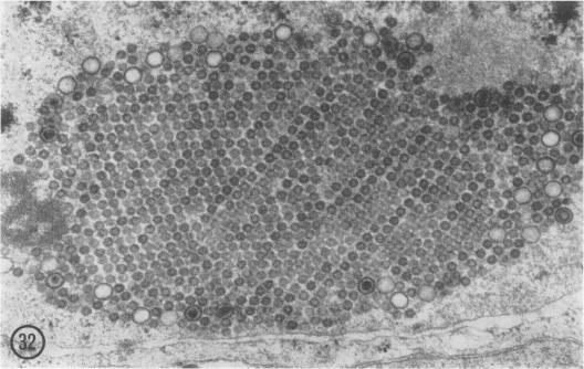
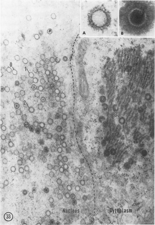
Development of the herpes-type virus of the frog kidney tumor was investigated by electron microscopy and high-resolution autoradiography in eyechamber transplants of tumor maintained at 7.5 C for up to 27 weeks. Virus particles were first detected at 10 weeks in nuclei containing aggregates of dense granular material. The initial incorporation of a pulse of (3)H-thymidine into these aggregates indicated that they contained newly synthesized viral deoxyribonucleic acid. Capsids enclosing doubleshelled cores were labeled with (3)H-thymidine before capsids with dense cores, and intermediate core forms were observed, suggesting that the double-shelled core transforms into the dense core. Particles with dense cores were observed while being enveloped by budding through the inner membrane of the nuclear envelope, and subsequently while being unenveloped in passing through the outer membrane into the cytoplasm. Virus particles within the cytoplasm acquired fibrillar coats and budded into vesicles, from which they were released, in enveloped form, at the cell surface. Tubular forms and particles considerably smaller than virus particles were regularly encountered in infected nuclei, and the relationship of these forms to virus replication is discussed.
通过电子显微镜和高分辨率放射自显影技术,对保存在7.5摄氏度长达27周的蛙肾肿瘤眼房移植瘤中疱疹型病毒的发育情况进行了研究。在含有致密颗粒物质聚集体的细胞核中,于第10周首次检测到病毒颗粒。将脉冲(3)H-胸腺嘧啶核苷最初掺入这些聚集体表明它们含有新合成的病毒脱氧核糖核酸。包裹双层核心的衣壳在包裹致密核心的衣壳之前被(3)H-胸腺嘧啶核苷标记,并且观察到中间核心形式,这表明双层核心转变为致密核心。观察到具有致密核心的颗粒在通过核膜内膜出芽时被包膜,随后在穿过外膜进入细胞质时被去包膜。细胞质内的病毒颗粒获得纤维状包膜并出芽进入囊泡,它们以包膜形式从囊泡中在细胞表面释放。在受感染的细胞核中经常遇到管状形式和比病毒颗粒小得多的颗粒,并讨论了这些形式与病毒复制的关系。