• 文献检索
  • 文档翻译
  • 深度研究
  • 学术资讯
  • Suppr Zotero 插件Zotero 插件
  • 邀请有礼
  • 套餐&价格
  • 历史记录
应用&插件
Suppr Zotero 插件Zotero 插件浏览器插件Mac 客户端Windows 客户端微信小程序
定价
高级版会员购买积分包购买API积分包
服务
文献检索文档翻译深度研究API 文档MCP 服务
关于我们
关于 Suppr公司介绍联系我们用户协议隐私条款
关注我们

Suppr 超能文献

核心技术专利:CN118964589B侵权必究
粤ICP备2023148730 号-1Suppr @ 2026

文献检索

告别复杂PubMed语法,用中文像聊天一样搜索,搜遍4000万医学文献。AI智能推荐,让科研检索更轻松。

立即免费搜索

文件翻译

保留排版,准确专业,支持PDF/Word/PPT等文件格式,支持 12+语言互译。

免费翻译文档

深度研究

AI帮你快速写综述,25分钟生成高质量综述,智能提取关键信息,辅助科研写作。

立即免费体验

视网膜色素上皮撕裂的发病机制。

Pathogenesis of tears of the retinal pigment epithelium.

作者信息

Gass J D

出版信息

Br J Ophthalmol. 1984 Aug;68(8):513-9. doi: 10.1136/bjo.68.8.513.

DOI:10.1136/bjo.68.8.513
PMID:6204685
原文链接:https://pmc.ncbi.nlm.nih.gov/articles/PMC1040403/
Abstract

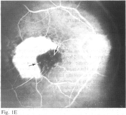
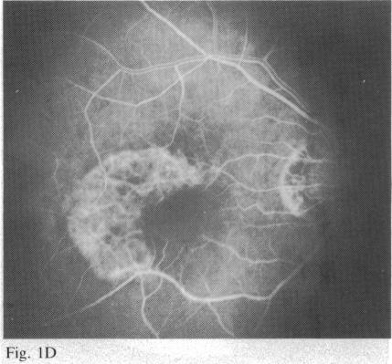
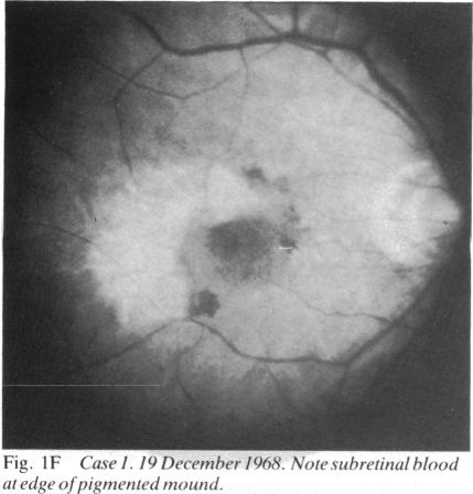
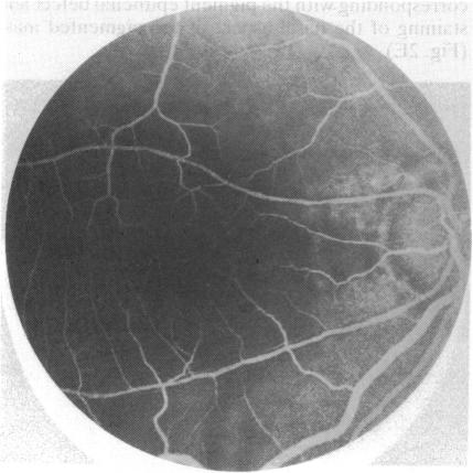
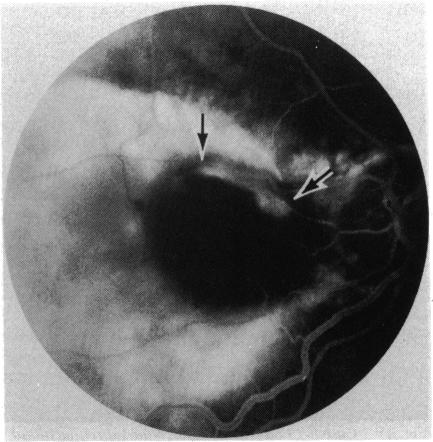
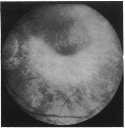
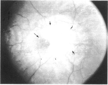
This report confirms a previous report that elderly patients with serous detachments of the pigment epithelium prior to and after developing a pigment epithelial tear at one border present a characteristic ophthalmoscopic and fluorescein angiographic appearance. Evidence is presented that subpigment epithelial choroidal neovascularisation and not irregular separation of the basement membrane from its pigment epithelium is the primary cause of the detachment and tear in the pigment epithelium.

摘要

本报告证实了之前的一份报告,即在色素上皮撕裂发生前后,出现色素上皮浆液性脱离的老年患者呈现出特征性的检眼镜和荧光素血管造影表现。有证据表明,色素上皮下脉络膜新生血管形成而非基底膜与其色素上皮的不规则分离是色素上皮脱离和撕裂的主要原因。

https://cdn.ncbi.nlm.nih.gov/pmc/blobs/c5ca/1040403/94f712dcba36/brjopthal00152-0015-b.jpg
https://cdn.ncbi.nlm.nih.gov/pmc/blobs/c5ca/1040403/e5b5f3a95b0f/brjopthal00152-0011-a.jpg
https://cdn.ncbi.nlm.nih.gov/pmc/blobs/c5ca/1040403/ffe984d4241a/brjopthal00152-0012-a.jpg
https://cdn.ncbi.nlm.nih.gov/pmc/blobs/c5ca/1040403/6216ebaa2bf3/brjopthal00152-0012-b.jpg
https://cdn.ncbi.nlm.nih.gov/pmc/blobs/c5ca/1040403/3f4341e590da/brjopthal00152-0012-c.jpg
https://cdn.ncbi.nlm.nih.gov/pmc/blobs/c5ca/1040403/db8922a859ec/brjopthal00152-0012-d.jpg
https://cdn.ncbi.nlm.nih.gov/pmc/blobs/c5ca/1040403/fff29ef133bc/brjopthal00152-0013-a.jpg
https://cdn.ncbi.nlm.nih.gov/pmc/blobs/c5ca/1040403/bd5fa83e219f/brjopthal00152-0013-b.jpg
https://cdn.ncbi.nlm.nih.gov/pmc/blobs/c5ca/1040403/4bf00f8a7759/brjopthal00152-0013-c.jpg
https://cdn.ncbi.nlm.nih.gov/pmc/blobs/c5ca/1040403/ea74b800446f/brjopthal00152-0013-d.jpg
https://cdn.ncbi.nlm.nih.gov/pmc/blobs/c5ca/1040403/966717511f2a/brjopthal00152-0014-a.jpg
https://cdn.ncbi.nlm.nih.gov/pmc/blobs/c5ca/1040403/a56a95a164d4/brjopthal00152-0014-b.jpg
https://cdn.ncbi.nlm.nih.gov/pmc/blobs/c5ca/1040403/6fce35387b5a/brjopthal00152-0014-c.jpg
https://cdn.ncbi.nlm.nih.gov/pmc/blobs/c5ca/1040403/1950b92b6a4b/brjopthal00152-0014-d.jpg
https://cdn.ncbi.nlm.nih.gov/pmc/blobs/c5ca/1040403/5ded39f8ebd8/brjopthal00152-0015-a.jpg
https://cdn.ncbi.nlm.nih.gov/pmc/blobs/c5ca/1040403/94f712dcba36/brjopthal00152-0015-b.jpg

相似文献

1
Pathogenesis of tears of the retinal pigment epithelium.视网膜色素上皮撕裂的发病机制。
Br J Ophthalmol. 1984 Aug;68(8):513-9. doi: 10.1136/bjo.68.8.513.
2
Clinicopathologic correlation of spontaneous retinal pigment epithelial tears with choroidal neovascular membranes in age-related macular degeneration.年龄相关性黄斑变性中自发性视网膜色素上皮撕裂与脉络膜新生血管膜的临床病理相关性
Ophthalmology. 1995 Feb;102(2):272-7. doi: 10.1016/s0161-6420(95)31045-7.
3
Giant pigment epithelial tear and exudative retinal detachment complicating choroidal effusions.巨大色素上皮撕裂及渗出性视网膜脱离并发脉络膜积液
Am J Ophthalmol. 1998 Sep;126(3):448-50. doi: 10.1016/s0002-9394(98)00104-4.
4
Indocyanine green angiography in retinal pigment epithelial tear.视网膜色素上皮撕裂的吲哚菁绿血管造影
Acta Ophthalmol Scand. 1998 Jun;76(3):384-5. doi: 10.1034/j.1600-0420.1998.760331.x.
5
Clinicopathological correlation of retinal pigment epithelial tears in exudative age related macular degeneration: pretear, tear, and scarred tear.渗出性年龄相关性黄斑变性中视网膜色素上皮撕裂的临床病理相关性:撕裂前、撕裂时及撕裂瘢痕期
Br J Ophthalmol. 2001 Apr;85(4):454-60. doi: 10.1136/bjo.85.4.454.
6
Serous retinal pigment epithelial detachment with a notch. A sign of occult choroidal neovascularization.伴有切迹的浆液性视网膜色素上皮脱离。隐匿性脉络膜新生血管的一个体征。
Retina. 1984 Fall-Winter;4(4):205-20. doi: 10.1097/00006982-198400440-00001.
7
Acute retinal pigment epithelial tear after photodynamic therapy.光动力治疗后急性视网膜色素上皮撕裂
Retina. 2001;21(6):661-5. doi: 10.1097/00006982-200112000-00017.
8
Tear of the retinal pigment epithelium and serous retinal detachment in a case of IgA nephropathy after renal transplantation.肾移植后IgA肾病患者出现视网膜色素上皮撕裂和浆液性视网膜脱离
Arch Ophthalmol. 2000 Apr;118(4):582-3.
9
Spontaneous rip of the retinal pigment epithelium with a macular hole in neovascular age-related macular degeneration.新生血管性年龄相关性黄斑变性中伴有黄斑裂孔的视网膜色素上皮自发撕裂
Am J Ophthalmol. 2005 Aug;140(2):316-8. doi: 10.1016/j.ajo.2005.01.028.
10
Prediction of retinal pigment epithelial tear in serous vascularized pigment epithelium detachment.预测浆液性血管性色素上皮脱离中的视网膜色素上皮撕裂。
Acta Ophthalmol. 2014 Feb;92(1):e50-6. doi: 10.1111/aos.12234. Epub 2013 Jul 2.

引用本文的文献

1
An atypical case of retinal pigment epithelium tear with remodeling and visual preservation.一例伴有重塑和视力保留的非典型视网膜色素上皮撕裂病例。
Taiwan J Ophthalmol. 2024 Oct 29;14(4):614-618. doi: 10.4103/tjo.TJO-D-24-00051. eCollection 2024 Oct-Dec.
2
Clinical Characteristics of Eyes with Neovascular Age-Related Macular Degeneration and Retinal Pigment Epithelium Tears.新生血管性年龄相关性黄斑变性合并视网膜色素上皮撕裂眼的临床特征
J Clin Med. 2023 Aug 24;12(17):5496. doi: 10.3390/jcm12175496.
3
Giant Retinal Pigment Epithelium Tear Resulting in Neurosensory Retinal Detachment: An Atypical Case.

本文引用的文献

1
Tears of detached retinal pigment epithelium.视网膜色素上皮脱离性裂孔
Br J Ophthalmol. 1981 Jun;65(6):417-22. doi: 10.1136/bjo.65.6.417.
2
A new perspective on Bruch's membrane and the retinal pigment epithelium.关于布鲁赫膜和视网膜色素上皮的新观点。
Br J Ophthalmol. 1982 Jan;66(1):17-25. doi: 10.1136/bjo.66.1.17.
3
Radial chorioretinal folds. A sign of choroidal neovascularization.视网膜脉络膜放射状皱褶。脉络膜新生血管的一个体征。
导致神经感觉性视网膜脱离的巨大视网膜色素上皮撕裂:1例非典型病例
J Vitreoretin Dis. 2022 Mar 1;6(6):461-466. doi: 10.1177/24741264211062928. eCollection 2022 Nov-Dec.
4
Fundus Autofluorescence and Clinical Applications.眼底自发荧光及其临床应用
J Ophthalmic Vis Res. 2021 Jul 29;16(3):432-461. doi: 10.18502/jovr.v16i3.9439. eCollection 2021 Jul-Sep.
5
Histopathology of Age-Related Macular Degeneration and Implications for Pathogenesis and Therapy.年龄相关性黄斑变性的组织病理学及其对发病机制和治疗的影响。
Adv Exp Med Biol. 2021;1256:67-88. doi: 10.1007/978-3-030-66014-7_3.
6
Optical coherence tomography features of the repair tissue following RPE tear and their correlation with visual outcomes.光相干断层扫描对 RPE 撕裂后修复组织的特征及其与视力结果的相关性。
Sci Rep. 2021 Mar 16;11(1):5962. doi: 10.1038/s41598-021-85270-x.
7
Changes in Treatment-Naive Pigment Epithelial Detachments Associated With the Initial Anti-Vascular Endothelial Growth Factor Injection: A Post Hoc Analysis From the HARBOR Trial.抗血管内皮生长因子治疗初始时未经治疗的色素上皮脱离的变化:HARBOR 试验的事后分析。
JAMA Ophthalmol. 2021 Feb 1;139(2):219-223. doi: 10.1001/jamaophthalmol.2020.5130.
8
Retinal Pigment Epithelial Detachment in Age-Related Macular Degeneration.年龄相关性黄斑变性中的视网膜色素上皮脱离
Ophthalmol Ther. 2020 Dec;9(4):739-756. doi: 10.1007/s40123-020-00291-5. Epub 2020 Aug 18.
9
Retinal Pigment Epithelium Tear.视网膜色素上皮撕裂。
Curr Ophthalmol Rep. 2015 Mar;3(1):26-33. doi: 10.1007/s40135-014-0057-1. Epub 2014 Dec 6.
10
Optical Coherence Tomography Angiography in Eyes with Non-infectious Posterior Uveitis; Some Practical Aspects.非感染性后葡萄膜炎患者眼中的光学相干断层扫描血管造影:一些实际问题
Med Hypothesis Discov Innov Ophthalmol. 2019 Winter;8(4):312-322. Epub 2019 Oct 1.
Arch Ophthalmol. 1981 Jun;99(6):1016-8. doi: 10.1001/archopht.1981.03930011016006.
4
Studies on the human macula. IV. Aging changes in Bruch's membrane.关于人类黄斑的研究。IV. 布鲁赫膜的衰老变化。
Arch Ophthalmol. 1967 Mar;77(3):410-20. doi: 10.1001/archopht.1967.00980020412022.
5
Photocoagulation in disciform macular degeneration.盘状黄斑变性中的光凝治疗。
Am J Ophthalmol. 1969 Jun;67(6):920-30. doi: 10.1016/0002-9394(69)90087-7.
6
Recent advances in the treatment of senile disciform macular degeneration by photocoagulation.光凝法治疗老年性盘状黄斑变性的最新进展。
Br J Ophthalmol. 1974 Apr;58(4):367-76. doi: 10.1136/bjo.58.4.367.
7
Serous detachments of the retinal pigment epithelium in patients with senile macular disease.老年性黄斑疾病患者视网膜色素上皮的浆液性脱离
Am J Ophthalmol. 1979 Oct;88(4):652-60. doi: 10.1016/0002-9394(79)90660-3.
8
Natural history of serous detachments of the retinal pigment epithelium.视网膜色素上皮浆液性脱离的自然病程。
Am J Ophthalmol. 1979 Oct;88(4):643-51. doi: 10.1016/0002-9394(79)90659-7.