• 文献检索
  • 文档翻译
  • 深度研究
  • 学术资讯
  • Suppr Zotero 插件Zotero 插件
  • 邀请有礼
  • 套餐&价格
  • 历史记录
应用&插件
Suppr Zotero 插件Zotero 插件浏览器插件Mac 客户端Windows 客户端微信小程序
定价
高级版会员购买积分包购买API积分包
服务
文献检索文档翻译深度研究API 文档MCP 服务
关于我们
关于 Suppr公司介绍联系我们用户协议隐私条款
关注我们

Suppr 超能文献

核心技术专利:CN118964589B侵权必究
粤ICP备2023148730 号-1Suppr @ 2026

文献检索

告别复杂PubMed语法,用中文像聊天一样搜索,搜遍4000万医学文献。AI智能推荐,让科研检索更轻松。

立即免费搜索

文件翻译

保留排版,准确专业,支持PDF/Word/PPT等文件格式,支持 12+语言互译。

免费翻译文档

深度研究

AI帮你快速写综述,25分钟生成高质量综述,智能提取关键信息,辅助科研写作。

立即免费体验

外阴上皮内瘤变及外阴微浸润癌

Vulvar intraepithelial neoplasia and microinvasive carcinoma of the vulva.

作者信息

Buckley C H, Butler E B, Fox H

出版信息

J Clin Pathol. 1984 Nov;37(11):1201-11. doi: 10.1136/jcp.37.11.1201.

DOI:10.1136/jcp.37.11.1201
PMID:6389601
原文链接:https://pmc.ncbi.nlm.nih.gov/articles/PMC498984/
Abstract

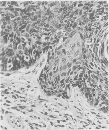
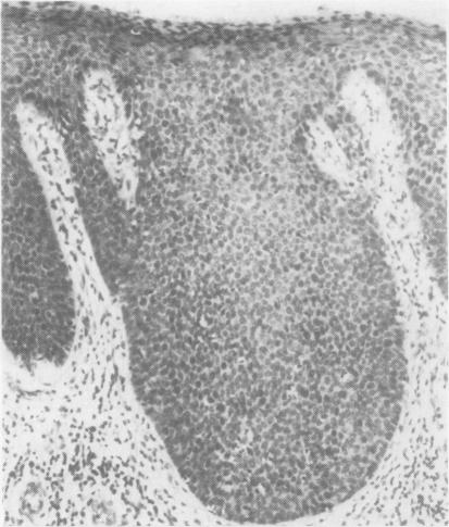
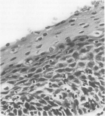
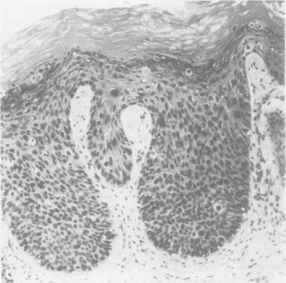
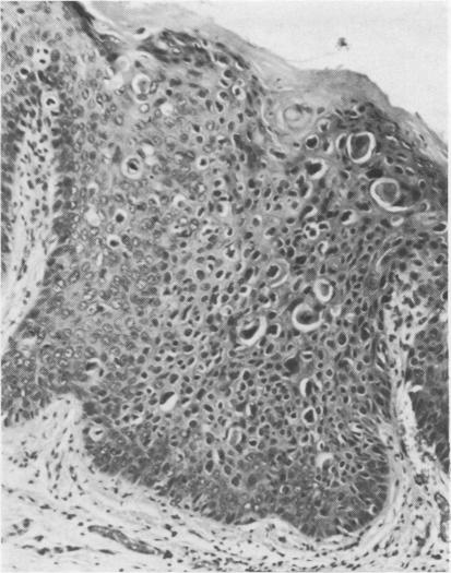
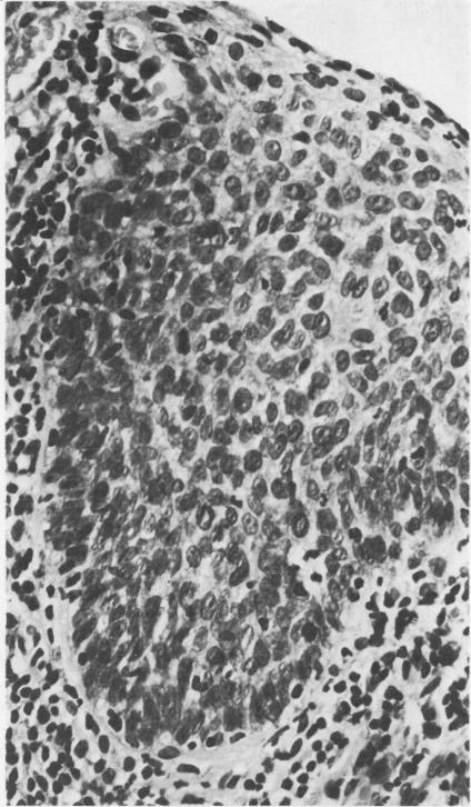
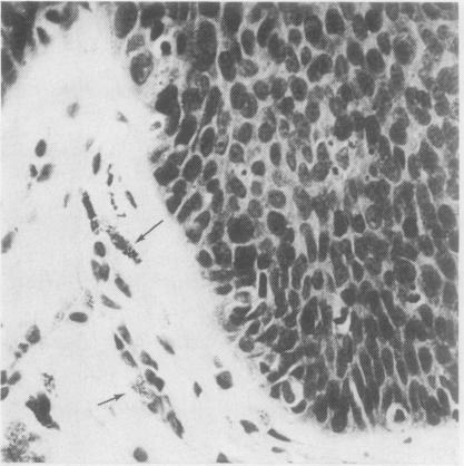
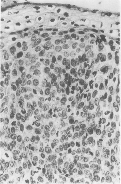
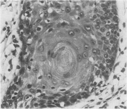
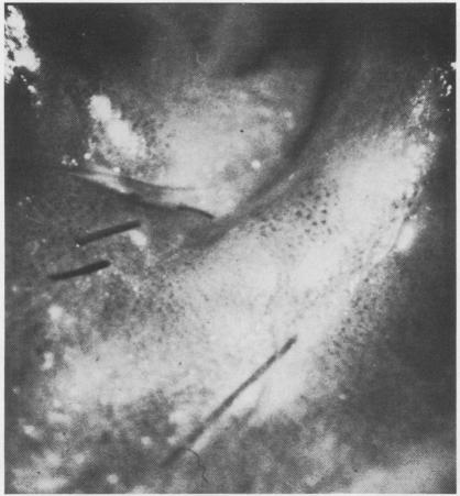
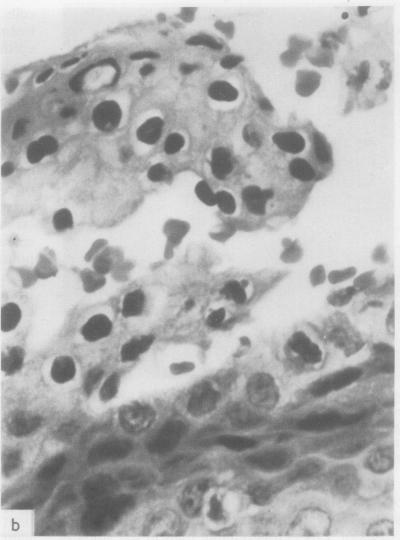
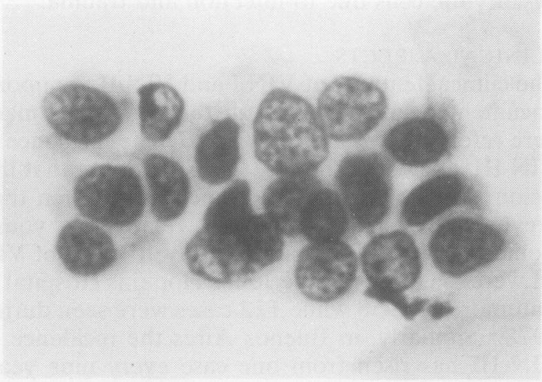
The pathological, cytological, and clinical features of vulvar intraepithelial neoplasia (VIN) are described. The rate of progression of VIN III to an invasive carcinoma is very low and spontaneous regression can occur. These features prevent the drawing of a direct analogy between vulvar and cervical intraepithelial neoplasia. The concept of microinvasive carcinoma of the vulva is discussed, and it is concluded that no satisfactory definition of this entity has been achieved.

摘要

本文描述了外阴上皮内瘤变(VIN)的病理、细胞学及临床特征。VIN III进展为浸润性癌的发生率很低,且可发生自发消退。这些特征使得外阴上皮内瘤变与宫颈上皮内瘤变无法直接进行类比。文中对外阴微浸润癌的概念进行了讨论,得出的结论是尚未对该实体达成令人满意的定义。

https://cdn.ncbi.nlm.nih.gov/pmc/blobs/7f6d/498984/25297445dd40/jclinpath00182-0010-a.jpg
https://cdn.ncbi.nlm.nih.gov/pmc/blobs/7f6d/498984/e82c08ae7e69/jclinpath00182-0002-a.jpg
https://cdn.ncbi.nlm.nih.gov/pmc/blobs/7f6d/498984/ee7e79172bf8/jclinpath00182-0002-b.jpg
https://cdn.ncbi.nlm.nih.gov/pmc/blobs/7f6d/498984/6f752273a024/jclinpath00182-0002-c.jpg
https://cdn.ncbi.nlm.nih.gov/pmc/blobs/7f6d/498984/69725bafa164/jclinpath00182-0002-d.jpg
https://cdn.ncbi.nlm.nih.gov/pmc/blobs/7f6d/498984/9aac689f076c/jclinpath00182-0003-a.jpg
https://cdn.ncbi.nlm.nih.gov/pmc/blobs/7f6d/498984/7a5dbf7d6758/jclinpath00182-0003-b.jpg
https://cdn.ncbi.nlm.nih.gov/pmc/blobs/7f6d/498984/122e3541da82/jclinpath00182-0004-a.jpg
https://cdn.ncbi.nlm.nih.gov/pmc/blobs/7f6d/498984/25049392dbf4/jclinpath00182-0004-b.jpg
https://cdn.ncbi.nlm.nih.gov/pmc/blobs/7f6d/498984/2f01801eb83a/jclinpath00182-0004-c.jpg
https://cdn.ncbi.nlm.nih.gov/pmc/blobs/7f6d/498984/b2e4eab675c4/jclinpath00182-0005-a.jpg
https://cdn.ncbi.nlm.nih.gov/pmc/blobs/7f6d/498984/68196c6a2c29/jclinpath00182-0006-a.jpg
https://cdn.ncbi.nlm.nih.gov/pmc/blobs/7f6d/498984/42c39d79ecae/jclinpath00182-0006-b.jpg
https://cdn.ncbi.nlm.nih.gov/pmc/blobs/7f6d/498984/606c499d8912/jclinpath00182-0006-c.jpg
https://cdn.ncbi.nlm.nih.gov/pmc/blobs/7f6d/498984/ab747e124c9d/jclinpath00182-0009-a.jpg
https://cdn.ncbi.nlm.nih.gov/pmc/blobs/7f6d/498984/25297445dd40/jclinpath00182-0010-a.jpg

相似文献

1
Vulvar intraepithelial neoplasia and microinvasive carcinoma of the vulva.外阴上皮内瘤变及外阴微浸润癌
J Clin Pathol. 1984 Nov;37(11):1201-11. doi: 10.1136/jcp.37.11.1201.
2
High-grade vulval intraepithelial neoplasia (VIN 3): a retrospective analysis of patient characteristics, management, outcome and relationship to squamous cell carcinoma of the vulva 1989-1999.高级别外阴上皮内瘤变(VIN 3):1989 - 1999年患者特征、治疗、结局及与外阴鳞状细胞癌关系的回顾性分析
Aust N Z J Obstet Gynaecol. 2002 Feb;42(1):69-74. doi: 10.1111/j.0004-8666.2002.00075.x.
3
Vulvar intraepithelial neoplasia with superficially invasive carcinoma of the vulva.外阴上皮内瘤变伴外阴浅表浸润性癌。
Br J Obstet Gynaecol. 1996 May;103(5):453-6. doi: 10.1111/j.1471-0528.1996.tb09772.x.
4
Vulvar intraepithelial neoplasia (severe atypia and carcinoma in situ). A clinicopathologic analysis of 41 cases.外阴上皮内瘤变(重度异型增生和原位癌)。41例临床病理分析。
Cancer. 1984 Oct 1;54(7):1429-34. doi: 10.1002/1097-0142(19841001)54:7<1429::aid-cncr2820540733>3.0.co;2-g.
5
Histopathologic study of thin vulvar squamous cell carcinomas and associated cutaneous lesions: a correlative study of 48 tumors in 44 patients with analysis of adjacent vulvar intraepithelial neoplasia types and lichen sclerosus.薄型外阴鳞状细胞癌及相关皮肤病变的组织病理学研究:44例患者48个肿瘤的相关性研究,分析相邻外阴上皮内瘤变类型和硬化性苔藓。
Am J Surg Pathol. 2006 Mar;30(3):310-8. doi: 10.1097/01.pas.0000180444.71775.1a.
6
Intraepithelial and invasive neoplasia of the vulva in association with human papillomavirus infection.与人类乳头瘤病毒感染相关的外阴上皮内瘤变和浸润性肿瘤
J Reprod Med. 1988 Jun;33(6):503-9.
7
Intraepithelial neoplasia of the vulva.外阴上皮内瘤变
Gynecol Oncol. 1995 Jan;56(1):8-21. doi: 10.1006/gyno.1995.1003.
8
Vulvar intraepithelial neoplasia in adolescents with abnormal Pap smear results: a series report.巴氏涂片结果异常的青少年外阴上皮内瘤变:系列报告
J Pediatr Adolesc Gynecol. 2004 Feb;17(1):45-8. doi: 10.1016/j.jpag.2003.11.001.
9
Vulvar intraepithelial neoplasia and carcinoma.外阴上皮内瘤变和癌。
Semin Cutan Med Surg. 1998 Sep;17(3):205-12. doi: 10.1016/s1085-5629(98)80015-1.
10
Vulvar intraepithelial neoplasia III: a clinical study of the outcome in 113 cases with relation to the later development of invasive vulvar carcinoma.外阴上皮内瘤变III级:113例患者结局与后续侵袭性外阴癌发生情况的临床研究
Obstet Gynecol. 1994 Nov;84(5):741-5.

引用本文的文献

1
Minichromosome maintenance (Mcm) proteins, cyclin B1 and D1, phosphohistone H3 and in situ DNA replication for functional analysis of vulval intraepithelial neoplasia.
Br J Cancer. 2003 Jan 27;88(2):257-62. doi: 10.1038/sj.bjc.6600729.

本文引用的文献

1
Intraepithelial carcinoma of the vulva.
Am J Obstet Gynecol. 1953 Mar;65(3):539-49. doi: 10.1016/0002-9378(83)90610-5.
2
Individualized treatment of stage I carcinoma of the vulva.外阴I期癌的个体化治疗
Obstet Gynecol. 1981 Jan;57(1):85-9.
3
Progressive histobiologic alterations in the development of vulvar cancer.外阴癌发展过程中的渐进性组织生物学改变。
Am J Obstet Gynecol. 1980 Sep 15;138(2):146-50. doi: 10.1016/0002-9378(80)90025-3.
4
Microinvasive carcinoma of the vulva: a clinicopathologic evaluation.外阴微小浸润癌:临床病理评估
Obstet Gynecol. 1980 Aug;56(2):234-8.
5
Carcinoma in situ of the vulva: a continuing challenge.外阴原位癌:一项持续的挑战。
Am J Obstet Gynecol. 1980 Apr 1;136(7):830-43. doi: 10.1016/0002-9378(80)91039-x.
6
Carcinoma in situ of the vulva.外阴原位癌
Obstet Gynecol. 1980 Feb;55(2):225-30.
7
Early invasive carcinoma of the vulva.早期外阴浸润癌
Am J Obstet Gynecol. 1981 Jul 1;140(5):563-9. doi: 10.1016/0002-9378(81)90233-7.
8
Microinvasive carcinoma of the vulva. A report of two cases with regional lymph node metastasis.外阴微浸润癌。两例伴有区域淋巴结转移的报告。
Diagn Gynecol Obstet. 1981 Spring;3(1):75-80.
9
Microinvasive carcinoma of the vulva.外阴微小浸润癌
Int J Gynecol Pathol. 1982;1(1):29-39. doi: 10.1097/00004347-198201000-00005.
10
Vulvar carcinoma in situ: a report of 28 cases.外阴原位癌:28例报告
Gynecol Oncol. 1982 Oct;14(2):236-42. doi: 10.1016/0090-8258(82)90096-8.