Tryphonas M, King D P, Jones P P
Proc Natl Acad Sci U S A. 1983 Mar;80(5):1445-8. doi: 10.1073/pnas.80.5.1445.
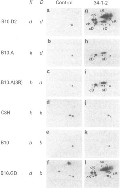
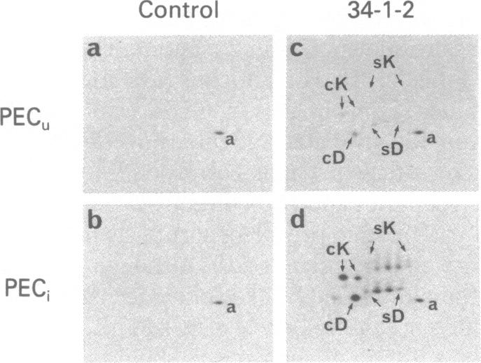
Immunoprecipitates obtained from [35S]methionine-labeled spleen cells by using monoclonal antibodies specific for H-2Kd and H-2Dd have been separated by two-dimensional polyacrylamide gel electrophoresis. Analysis of these gel patterns revealed the presence of an additional product of the K end of the H-2d complex, designated here as H-2K'. To determine whether H-2K' is a unique protein or a differentially glycosylated form of the previously characterized H-2Kd histocompatibility antigen, nonglycosylated molecules labeled in the presence of tunicamycin were examined. The results showed that both H-2K and H-2K' have distinct nonglycosylated polypeptide precursor forms. The approximate molecular weight differences between the fully glycosylated and nonglycosylated molecules also indicated the presence of three oligosaccharide side chains on H-2K', as is the case with H-2Kd, whereas H-2Dd has only two oligosaccharide units. The cellular expression of H-2K' was also investigated. Comparison of H-2 antigens immunoprecipitated from normal spleen cells and from thioglycollate-induced adherent peritoneal exudate cells cultured in the presence or absence of supernatant fluids from concanavalin A-stimulated spleen cells revealed that H-2K' was not expressed on the adherent peritoneal cells. This indicates that H-2K' is expressed in a tissue-specific manner, unlike the classical histocompatibility antigens H-2K and H-2D.
通过使用针对H-2Kd和H-2Dd的单克隆抗体,从[35S]甲硫氨酸标记的脾细胞中获得的免疫沉淀物,已通过二维聚丙烯酰胺凝胶电泳进行了分离。对这些凝胶图谱的分析揭示了H-2d复合体K端存在一种额外的产物,在此处命名为H-2K'。为了确定H-2K'是一种独特的蛋白质,还是先前已鉴定的H-2Kd组织相容性抗原的差异糖基化形式,对在衣霉素存在下标记的非糖基化分子进行了检测。结果表明,H-2K和H-2K'都有不同的非糖基化多肽前体形式。完全糖基化和非糖基化分子之间的近似分子量差异也表明,H-2K'上存在三条寡糖侧链,H-2Kd也是如此,而H-2Dd只有两条寡糖单元。还研究了H-2K'的细胞表达情况。比较从正常脾细胞以及在有无伴刀豆球蛋白A刺激的脾细胞上清液存在的情况下培养的巯基乙酸盐诱导的贴壁腹膜渗出细胞中免疫沉淀的H-2抗原,发现H-2K'在贴壁腹膜细胞上不表达。这表明H-2K'以组织特异性方式表达,这与经典组织相容性抗原H-2K和H-2D不同。