Thylefors B, Tønjum A M
Br J Ophthalmol. 1978 Jul;62(7):462-7. doi: 10.1136/bjo.62.7.462.
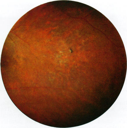
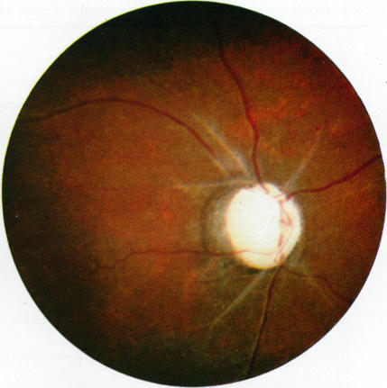
Lesions in the posterior segment of the eye in onchocerciasis may give visual field defects, but so far no detailed investigation has been done to determine the functional visual loss. Examination of the visual fields in 18 selected cases of onchocerciasis by means of a tangent screen test revealed important visual field defects associated with lesions in the posterior segment of the eye. Involvement of the optic nerve seemed to be important, giving rise to severely constricted visual fields. Cases of postneuritic optic atrophy showed a very uniform pattern of almost completely constricted visual fields, with only 5 to 10 degree central rest spared. Papillitis gave a similar severe constriction of the visual fields. The pattern of visual fields associated with optic neuropathy in onchocerciasis indicates that a progressive lesion of the optic nerve from the periphery may be responsible for the loss of vision. The visual field defects in onchocerciasis constitute a serious handicap, which must be taken into consideration when estimating the socioeconomic importance of the disease.
盘尾丝虫病患者眼后段的病变可能导致视野缺损,但迄今为止尚未进行详细调查以确定功能性视力丧失情况。通过切线屏试验对18例选定的盘尾丝虫病患者进行视野检查,发现重要的视野缺损与眼后段病变有关。视神经受累似乎很重要,导致视野严重缩窄。神经炎后视神经萎缩病例显示出几乎完全缩窄的视野非常一致的模式,仅保留5至10度的中央视野。视乳头炎也导致类似的严重视野缩窄。盘尾丝虫病中与视神经病变相关的视野模式表明,视神经从周边开始的进行性病变可能是视力丧失的原因。盘尾丝虫病的视野缺损构成严重障碍,在评估该疾病的社会经济重要性时必须予以考虑。