Klassen J, Milgrom F M, McCluskey R T
Am J Pathol. 1977 Jul;88(1):135-44.
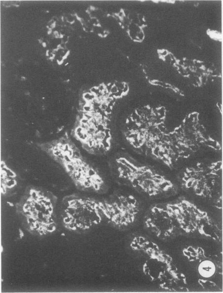
Rabbits injected with nonglomerular components of rabbit kidney incorporated in Freund's complete adjuvant develop a lesion characterized a) extensive interstitial fibrosis, tubular degenerative changes, and sparse focal lymphocytic infiltrates; b) the deposition of IgG and C3 in a granular pattern along the basement membranes of proximal convoluted tubules; and c) functional tubular defects if the lesions are severe. The antibodies were eluted from kidneys with such lesions and labeled with fluorescein isothiocyanate. It was shown that these fluorescein-labeled eluates reacted with the corresponding antigens in the tubular deposits and also with the antigens present in the brush border and/or cytoplasm of the proximal tubules. The antigens are found in proximal tubules of the kidney but not in brain, lung, heart, liver, spleen, bowel, muscles, or urine. They appear to be soluble but may also be present in the plasma membrane.
将兔肾非肾小球成分与弗氏完全佐剂混合后注射到兔子体内,会引发一种病变,其特征为:a) 广泛的间质纤维化、肾小管退行性变化以及稀疏的局灶性淋巴细胞浸润;b) IgG和C3以颗粒状沿近端曲管基底膜沉积;c) 如果病变严重,会出现功能性肾小管缺陷。从有此类病变的肾脏中洗脱抗体,并用异硫氰酸荧光素进行标记。结果显示,这些荧光素标记的洗脱液与肾小管沉积物中的相应抗原发生反应,也与近端小管刷状缘和/或细胞质中存在的抗原发生反应。这些抗原存在于肾脏的近端小管中,但在脑、肺、心脏、肝脏、脾脏、肠道、肌肉或尿液中不存在。它们似乎是可溶的,但也可能存在于质膜中。