Beveridge T J, Koval S F
Appl Environ Microbiol. 1981 Aug;42(2):325-35. doi: 10.1128/aem.42.2.325-335.1981.
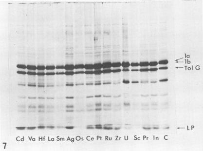
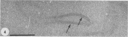
As representative of gram-negative bacteria, the isolated and purified envelopes of an Escherichia coli K-12 strain were used to determine metal-binding capacity. The envelopes were suspended in 5 mM metal solutions for 10 min and 23 degrees C, separated and washed by centrifugation, and analyzed for metal by either atomic absorption or X-ray fluorescence spectroscopy. Of 32 metals tested, large amounts (> 0.9 mumol/mg [dry weight]) of Hf and Os, intermediate amounts (0.1 to 0.4 mumol/mg [dry weight]) of Pb, Zn, Zr, Fe III, Mn, Mo, Mg, Co, and Ce IV, and small amounts (< 0.1 mumol/mg [dry weight]) of Na, K, Rb, Ca, Sr, Cu, Sc, La, Pr, Sm, U, Fe II, Ru, Ni, Hg, Pt, Pd, Au, and In were detected Li and V were not bound to the envelopes. Electron microscopy of unstained, thin-sectioned material provided an electron-scattering profile for localizing the bound metal within the envelope. Energy-dispersive X-ray analysis of thin sections detected all metals in single envelope vesicles. These data suggest that most metal deposition occurred at the polar head group regions of the constituent membranes or along the peptidoglycan layer. No leaching of envelope components was detected by monitoring radioactive probes within the lipopolysaccharide and peptidoglycan layers during metal uptake experiments, sodium dodecyl sulfate-polyacrylamide gel electrophoresis of proteins from metal-loaded envelopes, or protein and carbohydrate determinations on the wash fluids. These results suggest that membrane integrity was not disturbed under these ionic conditions.
作为革兰氏阴性菌的代表,使用大肠杆菌K-12菌株分离纯化的包膜来测定金属结合能力。将包膜悬浮于5 mM金属溶液中,在23℃下孵育10分钟,通过离心分离并洗涤,然后用原子吸收光谱法或X射线荧光光谱法分析金属含量。在测试的32种金属中,检测到大量(> 0.9 μmol/mg [干重])的铪(Hf)和锇(Os)、中等量(0.1至0.4 μmol/mg [干重])的铅(Pb)、锌(Zn)、锆(Zr)、铁(III)、锰(Mn)、钼(Mo)、镁(Mg)、钴(Co)和铈(IV),以及少量(< 0.1 μmol/mg [干重])的钠(Na)、钾(K)、铷(Rb)、钙(Ca)、锶(Sr)、铜(Cu)、钪(Sc)、镧(La)、镨(Pr)、钐(Sm)、铀(U)、铁(II)、钌(Ru)、镍(Ni)、汞(Hg)、铂(Pt)、钯(Pd)、金(Au)和铟(In);未检测到锂(Li)和钒(V)与包膜结合。对未染色的超薄切片材料进行电子显微镜观察,得到了用于在包膜内定位结合金属的电子散射图谱。对超薄切片进行能量色散X射线分析,在单个包膜小泡中检测到了所有金属。这些数据表明,大多数金属沉积发生在组成膜的极性头部基团区域或沿着肽聚糖层。在金属摄取实验期间,通过监测脂多糖和肽聚糖层内的放射性探针、对负载金属的包膜中的蛋白质进行十二烷基硫酸钠 - 聚丙烯酰胺凝胶电泳,或对洗涤液中的蛋白质和碳水化合物进行测定,均未检测到包膜成分的浸出。这些结果表明在这些离子条件下膜的完整性未受干扰。