• 文献检索
  • 文档翻译
  • 深度研究
  • 学术资讯
  • Suppr Zotero 插件Zotero 插件
  • 邀请有礼
  • 套餐&价格
  • 历史记录
应用&插件
Suppr Zotero 插件Zotero 插件浏览器插件Mac 客户端Windows 客户端微信小程序
定价
高级版会员购买积分包购买API积分包
服务
文献检索文档翻译深度研究API 文档MCP 服务
关于我们
关于 Suppr公司介绍联系我们用户协议隐私条款
关注我们

Suppr 超能文献

核心技术专利:CN118964589B侵权必究
粤ICP备2023148730 号-1Suppr @ 2026

文献检索

告别复杂PubMed语法,用中文像聊天一样搜索,搜遍4000万医学文献。AI智能推荐,让科研检索更轻松。

立即免费搜索

文件翻译

保留排版,准确专业,支持PDF/Word/PPT等文件格式,支持 12+语言互译。

免费翻译文档

深度研究

AI帮你快速写综述,25分钟生成高质量综述,智能提取关键信息,辅助科研写作。

立即免费体验

扁平苔藓样色素上皮病变与原田病。

Placoid pigment epitheliopathy and Harada's disease.

作者信息

Wright B E, Bird A C, Hamilton A M

出版信息

Br J Ophthalmol. 1978 Sep;62(9):609-21. doi: 10.1136/bjo.62.9.609.

DOI:10.1136/bjo.62.9.609
PMID:708678
原文链接:https://pmc.ncbi.nlm.nih.gov/articles/PMC1043308/
Abstract

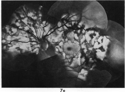
Twenty-six patients are described who suffered from acute bilateral multifocal pigment epithelial disease. In 7 the pattern of disease was indistinguishable from acute posterior multifocal placoid pigment epitheliopathy, while in 8 it was indistinguishable from Harada's disease. In a further 9 cases the pigment epithelial disease was associated with serious detachment of the retina simulating Harada's disease but without systemic symptoms; spontaneous resolution occurred within a few days, and there was no recurrence. One additional case had short-lived disease with detachment initially, but this was followed by severe recurrence, and the last patient had serious detachment in 1 eye but not the other. When seen as a whole these patients appeared to represent a continuous spectrum of disease making it difficult to define boundaries between one condition and another. The difficulties in distinguishing diseases according to morphology alone are emphasised.

摘要

本文描述了26例患有急性双侧多灶性色素上皮疾病的患者。其中7例疾病模式与急性后极部多灶性扁平色素上皮病变无法区分,8例与原田病无法区分。另外9例色素上皮疾病与类似原田病的严重视网膜脱离相关,但无全身症状;数天内自发消退,且无复发。另有1例最初有短暂性疾病伴视网膜脱离,但随后出现严重复发,最后1例患者1只眼有严重视网膜脱离,另1只眼则没有。总体来看,这些患者似乎代表了一系列连续的疾病,难以界定一种疾病与另一种疾病之间的界限。强调了仅根据形态学区分疾病的困难。

https://cdn.ncbi.nlm.nih.gov/pmc/blobs/651e/1043308/77897b705a1a/brjopthal00225-0043-b.jpg
https://cdn.ncbi.nlm.nih.gov/pmc/blobs/651e/1043308/e504590022f2/brjopthal00225-0033-a.jpg
https://cdn.ncbi.nlm.nih.gov/pmc/blobs/651e/1043308/21b02fdeb939/brjopthal00225-0033-b.jpg
https://cdn.ncbi.nlm.nih.gov/pmc/blobs/651e/1043308/45ba3b6eda8e/brjopthal00225-0033-c.jpg
https://cdn.ncbi.nlm.nih.gov/pmc/blobs/651e/1043308/a2e7a8a4f6a8/brjopthal00225-0034-a.jpg
https://cdn.ncbi.nlm.nih.gov/pmc/blobs/651e/1043308/23730d1e29dd/brjopthal00225-0034-b.jpg
https://cdn.ncbi.nlm.nih.gov/pmc/blobs/651e/1043308/683f0471caf4/brjopthal00225-0035-a.jpg
https://cdn.ncbi.nlm.nih.gov/pmc/blobs/651e/1043308/295dad3c356d/brjopthal00225-0035-b.jpg
https://cdn.ncbi.nlm.nih.gov/pmc/blobs/651e/1043308/c1d60552077b/brjopthal00225-0036-a.jpg
https://cdn.ncbi.nlm.nih.gov/pmc/blobs/651e/1043308/16067aaf415b/brjopthal00225-0036-b.jpg
https://cdn.ncbi.nlm.nih.gov/pmc/blobs/651e/1043308/a10a8d9ad46b/brjopthal00225-0037-a.jpg
https://cdn.ncbi.nlm.nih.gov/pmc/blobs/651e/1043308/fff8ce36a080/brjopthal00225-0038-a.jpg
https://cdn.ncbi.nlm.nih.gov/pmc/blobs/651e/1043308/2fa4e6ac1830/brjopthal00225-0038-b.jpg
https://cdn.ncbi.nlm.nih.gov/pmc/blobs/651e/1043308/8e7530870e43/brjopthal00225-0040-a.jpg
https://cdn.ncbi.nlm.nih.gov/pmc/blobs/651e/1043308/ff8da5bab7ab/brjopthal00225-0040-b.jpg
https://cdn.ncbi.nlm.nih.gov/pmc/blobs/651e/1043308/e7176c887b8d/brjopthal00225-0041-a.jpg
https://cdn.ncbi.nlm.nih.gov/pmc/blobs/651e/1043308/0e600ca0c84a/brjopthal00225-0041-b.jpg
https://cdn.ncbi.nlm.nih.gov/pmc/blobs/651e/1043308/d538522c2532/brjopthal00225-0042-a.jpg
https://cdn.ncbi.nlm.nih.gov/pmc/blobs/651e/1043308/d57e0e387aac/brjopthal00225-0043-a.jpg
https://cdn.ncbi.nlm.nih.gov/pmc/blobs/651e/1043308/77897b705a1a/brjopthal00225-0043-b.jpg

相似文献

1
Placoid pigment epitheliopathy and Harada's disease.扁平苔藓样色素上皮病变与原田病。
Br J Ophthalmol. 1978 Sep;62(9):609-21. doi: 10.1136/bjo.62.9.609.
2
Acute posterior multifocal placoid pigment epitheliopathy and Harada's disease.急性后极部多灶性扁平色素上皮病变与原田病。
Ann Ophthalmol. 1983 Jan;15(1):58-62.
3
[Angiographic signs of type III placoid epitheliopathy, Harada's disease and uveal effusion].
Bull Soc Ophtalmol Fr. 1989 Feb;89(2):331-5.
4
[On Harada's disease (author's transl)].关于原田病(作者译)
Klin Monbl Augenheilkd. 1978 Aug;173(2):243-6.
5
Atypical Acute Posterior Multifocal Placoid Pigment Epitheliopathy.非典型急性后极部多灶性扁平色素上皮病变
Optom Vis Sci. 2016 Jan;93(1):103-6. doi: 10.1097/OPX.0000000000000756.
6
Harada's, Behcet's, Vogt-Koyanagi syndromes--are they clinical entities?原田氏病、白塞氏病、Vogt-小柳综合征——它们是临床实体吗?
Trans Am Acad Ophthalmol Otolaryngol. 1973 May-Jun;77(3):OP281-90.
7
Spectral domain optical coherence tomography and autofluorescence in a case of acute posterior multifocal placoid pigment epitheliopathy mimicking Vogt-Koyanagi-Harada disease: case report and review of literature.谱域光相干断层扫描和自发荧光在一例急性后多发性盘状色素上皮病变模仿 Vogt-Koyanagi-Harada 病的病例报告及文献复习。
Ocul Immunol Inflamm. 2011 Feb;19(1):42-7. doi: 10.3109/09273948.2010.521610. Epub 2010 Oct 31.
8
[Fluoro-angiographic study of Harada's disease].[原田病的荧光血管造影研究]
Bull Mem Soc Fr Ophtalmol. 1972;85(0):355-63.
9
[Uveoencephalitis (Harada's disease)].[葡萄膜脑炎(原田病)]
Oftalmol Zh. 1974;29(7):552-3.
10
[Harada's disease. Report of a case].[原田氏病。病例报告]
Med Trop (Mars). 1995;55(4 Pt 2):459-61.

引用本文的文献

1
Uveal vascular bed in health and disease: lesions produced by occlusion of the uveal vascular bed and acute uveal ischaemic lesions seen clinically. Paper 2 of 2.眼葡萄膜血管床在健康和疾病中的表现:眼葡萄膜血管闭塞引起的病变和临床上观察到的急性眼葡萄膜缺血性病变。全文 2 篇中的第 2 篇。
Eye (Lond). 2023 Sep;37(13):2617-2648. doi: 10.1038/s41433-023-02417-y. Epub 2023 Apr 25.
2
Multimodal imaging of the course of retinal changes in acute posterior multifocal placoid pigment epitheliopathy with bilateral retinal detachment: a case report.多模态成像在伴有双侧视网膜脱离的急性后部多灶性脉络膜视网膜炎中视网膜变化过程中的应用:一例报告。
BMC Ophthalmol. 2022 Oct 7;22(1):400. doi: 10.1186/s12886-022-02624-3.
3

本文引用的文献

1
VIRUSLIKE INCLUSION BODIES IN SUBRETINAL FLUID IN UVEO-ENCEPHALITIS.
Am J Ophthalmol. 1964 Dec;58:940-5. doi: 10.1016/0002-9394(64)90002-9.
2
Acute posterior multifocal placoid pigment epitheliopathy.急性后极部多灶性扁平色素上皮病变
Arch Ophthalmol. 1968 Aug;80(2):177-85. doi: 10.1001/archopht.1968.00980050179005.
3
Lymphocyte transformation test in sympathetic ophthalmitis and the Vogt-Koyanagi-Harada syndrome.交感性眼炎和Vogt-小柳-原田综合征中的淋巴细胞转化试验。
Br J Ophthalmol. 1971 Dec;55(12):850-2. doi: 10.1136/bjo.55.12.850.
Spectral domain optical coherence tomography as an adjunctive tool for screening Behçet uveitis.
频域光学相干断层扫描作为贝赫切特葡萄膜炎筛查的辅助工具。
PLoS One. 2018 Dec 11;13(12):e0208254. doi: 10.1371/journal.pone.0208254. eCollection 2018.
4
Uveitic macular edema.葡萄膜炎性黄斑水肿
Eye (Lond). 2016 Oct;30(10):1277-1292. doi: 10.1038/eye.2016.115. Epub 2016 Jun 3.
5
Subretinal fluid in acute posterior multifocal placoid pigment epitheliopathy.急性后极部多发性斑状脉络膜视网膜病变的视网膜下液。
Retina. 2010 May;30(5):810-4. doi: 10.1097/IAE.0b013e3181c596f8.
6
Acute posterior multifocal placoid pigment epitheliopathy.急性后极部多灶性鳞状色素上皮病变
Br J Ophthalmol. 1995 Apr;79(4):384-9. doi: 10.1136/bjo.79.4.384.
7
Macular lesions secondary to choroidal vascular disorders.
Int Ophthalmol. 1983 Feb;6(2):161-70. doi: 10.1007/BF00127645.
8
Acute occlusive disorders of the choroidal vasculature.
Int Ophthalmol. 1983 Feb;6(2):139-48. doi: 10.1007/BF00127642.
9
Multifocal and recurrent (serous) choroidopathy (MARC) syndrome: a new variety of idiopathic central serous choroidopathy.多灶性复发性(浆液性)脉络膜病变(MARC)综合征:特发性中心性浆液性脉络膜病变的一种新类型。
Doc Ophthalmol. 1984 Jan 30;56(3):203-35. doi: 10.1007/BF00159074.
10
Idiopathic sub-retinal neovascular membranes in the macula (hemorrhagic macular choroidopathy of young adults). Clinical report and effectiveness of laser treatment.
Doc Ophthalmol. 1986;64(3):311-43. doi: 10.1007/BF00161734.
4
Pigmentary epitheliopathy and erythema nodosum.
Arch Ophthalmol. 1971 Mar;85(3):369-72. doi: 10.1001/archopht.1971.00990050371025.
5
Acute retinal pigment epitheliitus.
Am J Ophthalmol. 1972 Aug;74(2):193-205. doi: 10.1016/0002-9394(72)90535-1.
6
Diverse manifestations of acute posterior multifocal placoid pigment epitheliopathy.急性后极部多灶性鳞状色素上皮病变的多样表现
Am J Ophthalmol. 1974 May;77(5):659-62. doi: 10.1016/0002-9394(74)90527-3.
7
Multifocal placoid pigment epitheliopathy.
Am J Ophthalmol. 1973 Oct;76(4):511-8. doi: 10.1016/0002-9394(73)90739-3.
8
Acute posterior multifocal placoid pigment epitheliopathy.急性后极部多灶性扁平色素上皮病变
Arch Ophthalmol. 1973 May;89(5):373-6. doi: 10.1001/archopht.1973.01000040375004.
9
Placoid pigment epitheliopathy. Presenting with bilateral serous retinal detachment.
Br J Ophthalmol. 1972 Dec;56(12):881-6. doi: 10.1136/bjo.56.12.881.
10
Placoid pigment epitheliopathy with retinal vasculitis and papillitis.伴有视网膜血管炎和视乳头炎的板层样色素上皮病变
Br J Ophthalmol. 1972 Dec;56(12):875-80. doi: 10.1136/bjo.56.12.875.