Egbert P R, Fajardo L F, Donaldson S S, Moazed K
Br J Ophthalmol. 1980 Sep;64(9):660-5. doi: 10.1136/bjo.64.9.660.
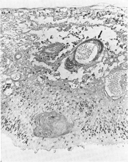
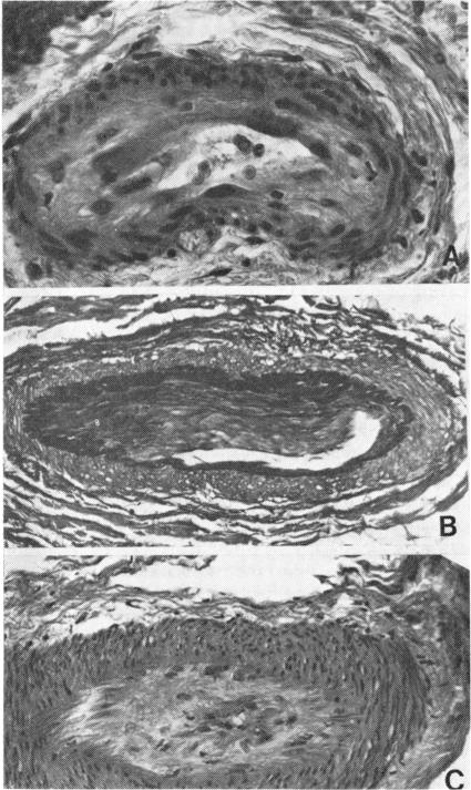
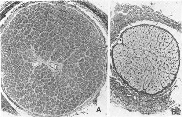
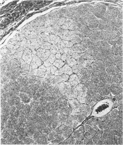
Radiation-induced ocular lesions in the posterior eye and orbit were investigated in 33 surgical specimens of patients with retinoblastoma. The eyes were obtained from children 7 months to 6 years of age. Seventeen eyes were irradiated; 16 eyes had not received irradiation and served as controls. The majority of the irradiated eyes were treated with 6000 rads of external beam radiation. They were removed at a mean of 23 months after radiotherapy. All specimens were examined simultaneously by 2 observers without knowledge of treatment and analysed for the presence or absence of 15 lesions. The most consistent lesions in the irradiated eyes were abnormalities of the retinal vessels (11 of 17 eyes) and striking changes in the ciliary arteries (13 of 17 eyes). The retinal vessels showed thickening of the wall, often caused by deposition of fibrillary material, sometimes with fibrin deposits. The most consistent lesion was myointimal proliferation with narrowing of the ciliary arteries. Lesions of the central retinal artery were less common but occurred only in irradiated patients.
对33例视网膜母细胞瘤患者的手术标本进行了研究,以观察后眼和眼眶的辐射性眼部病变。这些眼睛取自7个月至6岁的儿童。17只眼睛接受了放疗;16只眼睛未接受放疗作为对照。大多数接受放疗的眼睛接受了6000拉德的外照射。它们在放疗后平均23个月被摘除。所有标本由2名观察者在不知道治疗情况的前提下同时进行检查,并分析15种病变的有无。接受放疗的眼睛中最常见的病变是视网膜血管异常(17只眼中有11只)和睫状动脉显著改变(17只眼中有13只)。视网膜血管显示管壁增厚,通常是由纤维状物质沉积引起的,有时伴有纤维蛋白沉积。最常见的病变是肌内膜增生伴睫状动脉狭窄。视网膜中央动脉病变较少见,但仅发生在接受放疗的患者中。