Quinones M A, Young J B, Waggoner A D, Ostojic M C, Ribeiro L G, Miller R R
Br Heart J. 1980 Dec;44(6):612-20. doi: 10.1136/hrt.44.6.612.
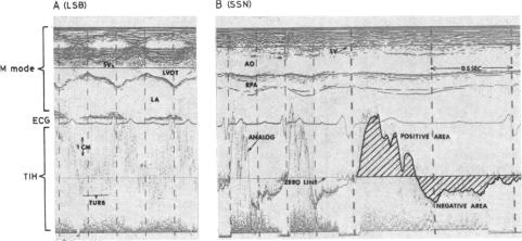
Pulsed Doppler echocardiography was employed to detect disturbed or turbulent flow diagnostic of aortic or mitral regurgitation. Sensitivity, specificity, diagnostic accuracy, and predictive value were assessed by the independent interpretation and comparison of aortic root angiograms (91 patients) and left ventriculograms (94 patients) to the time interval histogram display of the pulsed Doppler. Sensitivity of Doppler in detecting mitral regurgitation was 94 per cent, with specificity 89 per cent, predictive value 81 per cent, and diagnostic accuracy 90 per cent (32 patients with, 62 without regurgitation). In aortic regurgitation, sensitivity was also 94 per cent, specificity 82 per cent, predictive value 94 per cent, and the diagnostic accuracy was 91 per cent (69 patients with, 22 without aortic regurgitation). Additionally, no Doppler evidence of mitral or aortic regurgitation was present in 20 normal subjects. The aetiology of left-sided valvular regurgitation varied widely, with prosthetic valvular insufficiency being the cause of mitral and aortic regurgitation in seven and 10 patients, respectively. Sixteen of 17 (94%) paraprosthetic leaks were correctly identified by pulsed Doppler. In patients with aortic regurgitation the flow-velocity curve recorded in the ascending aorta frequently showed a negative (or reversed) diastolic component, the magnitude of which (expressed as percentage negative area) correlated significantly with angiographic severity of regurgitation. Thus, pulsed Doppler echocardiography is a highly accurate and objective non-invasive technique for detecting mitral and aortic regurgitation. In aortic regurgitation, estimation of severity is possible from inspection of the Doppler ascending aortic flow velocity curve.
采用脉冲多普勒超声心动图检测紊乱或湍流,以诊断主动脉瓣或二尖瓣反流。通过对91例患者的主动脉根部血管造影和94例患者的左心室造影进行独立解读,并与脉冲多普勒的时间间隔直方图显示进行比较,评估其敏感性、特异性、诊断准确性和预测价值。多普勒检测二尖瓣反流的敏感性为94%,特异性为89%,预测价值为81%,诊断准确性为90%(32例有反流,62例无反流)。在主动脉瓣反流中,敏感性也为94%,特异性为82%,预测价值为94%,诊断准确性为91%(69例有主动脉瓣反流,22例无主动脉瓣反流)。此外,20名正常受试者未发现二尖瓣或主动脉瓣反流的多普勒证据。左侧瓣膜反流的病因差异很大,人工瓣膜功能不全分别是7例二尖瓣反流和10例主动脉瓣反流的原因。17例人工瓣膜旁漏中有16例(94%)被脉冲多普勒正确识别。在主动脉瓣反流患者中,升主动脉记录的血流速度曲线经常显示舒张期成分呈负向(或反向),其大小(以负向面积百分比表示)与血管造影所示反流严重程度显著相关。因此,脉冲多普勒超声心动图是一种检测二尖瓣和主动脉瓣反流的高度准确且客观的非侵入性技术。对于主动脉瓣反流,通过检查多普勒升主动脉血流速度曲线可以估计反流严重程度。