Monroy A, Ridley D S, Heather C J, Ridley M J
Br J Exp Pathol. 1980 Dec;61(6):601-10.
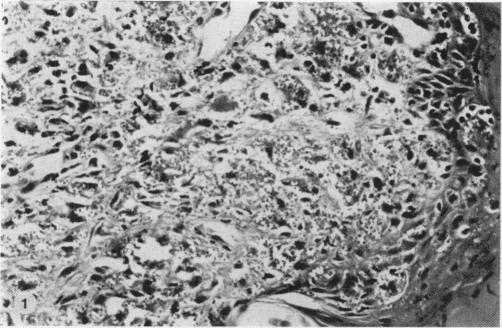
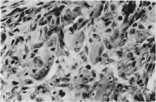
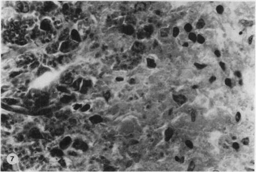
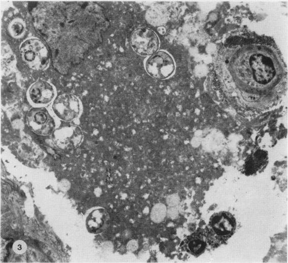
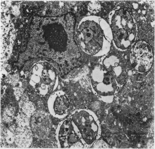
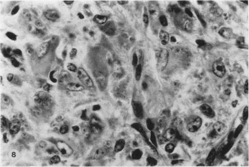
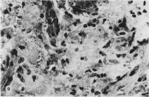
Nineteen guinea-pigs were each inoculated intradermally with 10(6) amastigotes of Leishmania enriettii, and the development of the lesions was followed from Weeks 4 to 10 with a view to elucidating the histological mechanisms involved with the elimination of parasites. Electron microscopic observations were made in 1 animal. Extensive necrosis of the parasite-laden macrophages was observed in 7 out of 7 animals at 4 and 5 weeks. In the ulcerated core of the lesion at 4 weeks no intact macrophages could be identified. Very many amastigotes were extracellular. Others were present in the cytoplasm of residual macrophages the cell walls of which had disintegrated. Necrosis was less marked at 8 weeks and absent in the resolving lesions at 10 weeks. Signs of stimulation or maturation of macrophages were only apparent when parasites were few. At 4 weeks macrophages were almost all of the non-stimulated form, but cytological evidence of activation became progressively more definite and widespread from 5 to 8 weeks, starting at the periphery of the lesion. Ultrastructural observations of amastigotes suggested that there might be more than one mechanism of degradation. It appeared that the majority of parasites were released through necrosis and discharged through the ulcer, and that intracellular degradation of the remaining parasites was important mainly in the later phase before resolution. The first phase was associated mainly with plasma-cell production, the second mainly with lymphocytes.
19只豚鼠每只皮内接种10^6个恩氏利什曼原虫无鞭毛体,从第4周开始观察病变发展直至第10周,以阐明与寄生虫清除相关的组织学机制。对1只动物进行了电子显微镜观察。在4周和5周时,7只动物中有7只观察到富含寄生虫的巨噬细胞广泛坏死。在4周时病变的溃疡核心部位,未发现完整的巨噬细胞。大量无鞭毛体位于细胞外。其他无鞭毛体存在于残余巨噬细胞的细胞质中,这些巨噬细胞的细胞壁已经解体。8周时坏死不太明显,10周时正在消退的病变中无坏死现象。只有当寄生虫数量很少时,巨噬细胞的刺激或成熟迹象才明显。4周时巨噬细胞几乎全是非刺激型,但从5周到8周,激活的细胞学证据从病变周边开始逐渐变得更加明确和广泛。对无鞭毛体的超微结构观察表明,可能存在不止一种降解机制。似乎大多数寄生虫通过坏死释放并通过溃疡排出,而剩余寄生虫的细胞内降解主要在消退前的后期阶段起重要作用。第一阶段主要与浆细胞产生有关,第二阶段主要与淋巴细胞有关。