Safran A B, Halfon A, Safran E, Mermoud C
Department of Ophthalmology, Geneva University Hospital, Switzerland.
Br J Ophthalmol. 1995 Feb;79(2):118-24. doi: 10.1136/bjo.79.2.118.
To determine principles which regulate the occurrence of angioscotomata in automated static perimetry, variations in light sensitivity were correlated with the location and diameter of neighbouring retinal vessels.
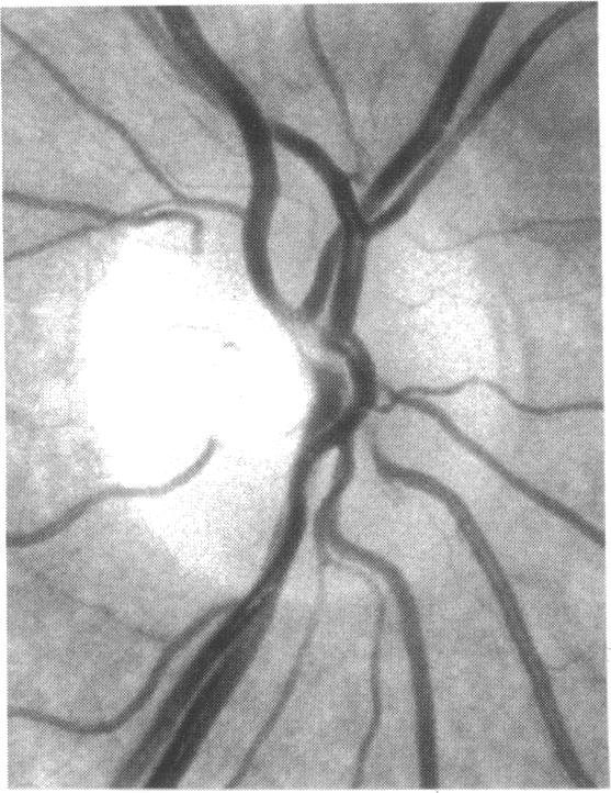
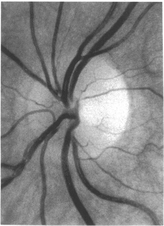
Ten normal eyes were tested with the Octopus 2000R, using a 0.431 degree light stimulus. Sensitivity was quantified in points located around the blind spot, according to a regular, 0.5 degree constant, grid pattern. From 336 to 443 locations were tested in each eye. The resulting printouts were superimposed on corresponding fundus photographs. At each tested point, the following five additional variables were evaluated: the diameters of the closest and the second closest vessel (in 0.1 degree units); the distances of the apparent location of the tested point to the closest and the second closest vessel (in 0.25 degree units); and the distance between the two closest vessels (in 0.25 degree units). Altogether, 3869 locations were tested and 23,214 values were quantified.
The following two conditions were found to be related to a reduction in sensitivity: (1) proximity (< 0.25 degree) to a large vessel (> or = 0.5 degree in diameter); (2) proximity (< 0.25 degree) to one of two adjacent (< 0.5 degree distant), moderately large vessels (0.3 degree to 0.4 degree in diameter). In condition 1, sensitivity was 51.3% and specificity was 92.2%; in condition 2, sensitivity was 16.2% and specificity was 98.3%; and with a combination of conditions 1 and 2, sensitivity was 67.6% and specificity was 90.5%. Increase by 0.1 degree of an adjacent vessel which was 0.4 degree in diameter markedly affected light sensitivity.
Modifications in vessel diameter are observed in a number of circumstances, including adaptive vascular response to changes in ambient conditions and obstructive disorders of retinal vessels. These findings indicate that changes in vessel diameter over time can result in fluctuation of sensitivity. It is concluded that, in contrast with what is commonly stated, when ocular media are unaltered and the subject's collaboration is adequate, temporal variations in measured thresholds do not necessarily reflect functional changes in nervous tissues in the visual pathways.
为确定自动静态视野检查中调节暗点出现的原理,将光敏感度变化与相邻视网膜血管的位置和直径相关联。
使用Octopus 2000R对10只正常眼睛进行检测,采用0.431度的光刺激。根据规则的、0.5度恒定的网格模式,在盲点周围的点上对敏感度进行量化。每只眼睛测试336至443个位置。将所得打印结果叠加在相应的眼底照片上。在每个测试点,评估以下五个额外变量:最接近和第二接近血管的直径(以0.1度为单位);测试点的表观位置到最接近和第二接近血管的距离(以0.25度为单位);以及两个最接近血管之间的距离(以0.25度为单位)。总共测试了3869个位置,量化了23214个值。
发现以下两种情况与敏感度降低有关:(1)靠近(<0.25度)大血管(直径≥0.5度);(2)靠近(<0.25度)两个相邻(距离<0.5度)、中等大小血管(直径0.3度至0.4度)之一。在情况1中,敏感度为51.3%,特异性为92.2%;在情况2中,敏感度为16.2%,特异性为98.3%;情况1和2组合时,敏感度为67.6%,特异性为90.5%。直径为0.4度的相邻血管直径增加0.1度会显著影响光敏感度。
在多种情况下观察到血管直径的改变,包括对环境条件变化的适应性血管反应和视网膜血管阻塞性疾病。这些发现表明血管直径随时间的变化可导致敏感度波动。得出结论,与通常所说的相反,当眼介质未改变且受试者配合良好时,测量阈值的时间变化不一定反映视觉通路中神经组织的功能变化。