Kimura W, Nagai H
First Department of Surgery, Faculty of Medicine, University of Tokyo, Japan.
Ann Surg. 1995 Apr;221(4):359-63. doi: 10.1097/00000658-199504000-00005.
The authors precisely examined the topography of the duodenum, pancreas, bile duct, and supplying vessels from the perspective of performing duodenum-preserving resection of the pancreatic head.
Little has been reported regarding the detailed surgical anatomy that is crucial in this procedure.
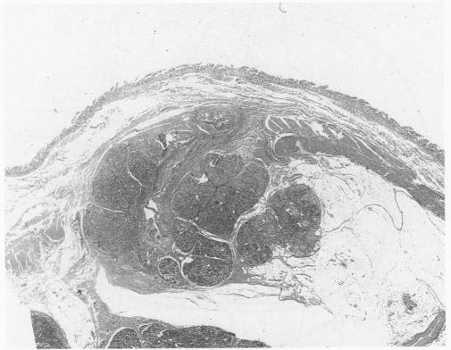
The authors precisely examined the local anatomy of the pancreas head and duodenum, using materials from 40 autopsy cases.
Arcade formation between the anterior superior pancreaticoduodenal (ASPD) artery and the anterior inferior pancreaticoduodenal (AIPD) artery was found in all of the cases. After departing from the gastroduodenal artery, the ASPD ran toward a point 1.5 cm below the papilla of Vater, then turned to the posterior aspect of the pancreas to joint the AIPD. In 88% of the cases, an arcade was found between the posterior superior pancreaticoduodenal (PSPD) artery and the posterior inferior pancreaticoduodenal (PIPD) artery. The ASPD, AIPD, PSPD, PIPD, or their branches to the duodenum, the bile duct, and the papilla of Vater were not completely buried in the pancreatic parenchyma in any of these cases. Generally, it was easy to dissect the pancreas from the duodenum because of the loose connection. Near the accessory papilla, however, dissection of the vessels was difficult, and the pancreatic parenchyma sometimes was found in the wall of the duodenum. Dissection of the pancreas from the common bile duct and identification of the main pancreatic duct at the junction with the terminal portion of the bile duct were straightforward in all cases.
It may be possible to remove the head of the pancreas while preserving of the vascular arcades and their branches to the duodenum, the bile duct, and the papilla of Vater.
作者从实施保留十二指肠的胰头切除术的角度,精确研究十二指肠、胰腺、胆管及供血血管的局部解剖结构。
关于此手术中至关重要的详细手术解剖学的报道较少。
作者使用40例尸检病例的材料,精确研究胰头和十二指肠的局部解剖结构。
所有病例均发现胰十二指肠上前(ASPD)动脉与胰十二指肠下前(AIPD)动脉之间形成动脉弓。ASPD自胃十二指肠动脉发出后,走向距 Vater 壶腹下方1.5 cm处,然后转向胰腺后方与 AIPD 汇合。88%的病例中,发现胰十二指肠上后(PSPD)动脉与胰十二指肠下后(PIPD)动脉之间存在动脉弓。在所有这些病例中,ASPD、AIPD、PSPD、PIPD 或它们至十二指肠、胆管及 Vater 壶腹的分支均未完全埋入胰腺实质。一般来说,由于二者连接疏松,胰腺与十二指肠易于分离。然而,在副乳头附近,血管分离困难,有时在十二指肠壁中可发现胰腺实质。在所有病例中,胰腺与胆总管的分离以及在胆总管终末部与主胰管交界处识别主胰管均很容易。
在保留血管弓及其至十二指肠、胆管和 Vater 壶腹的分支的同时,有可能切除胰头。