Whitley P, Zander T, Ehrmann M, Haardt M, Bremer E, von Heijne G
Department of Molecular Biology, Karolinska Institute Center for Structural Biochemistry, NOVUM, Huddinge, Sweden.
EMBO J. 1994 Oct 3;13(19):4653-61. doi: 10.1002/j.1460-2075.1994.tb06788.x.
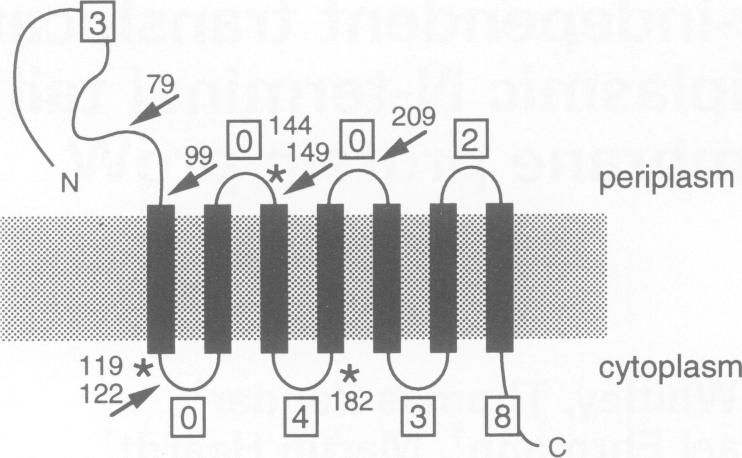
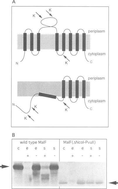
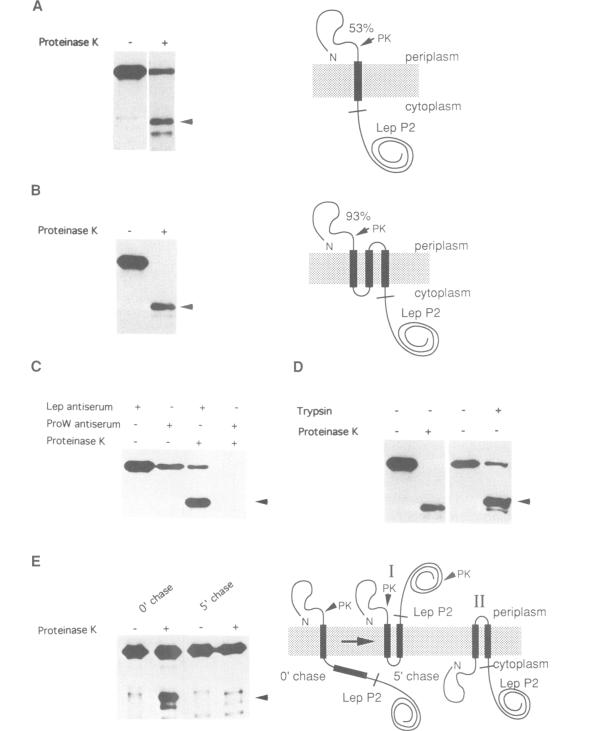
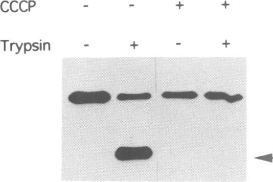
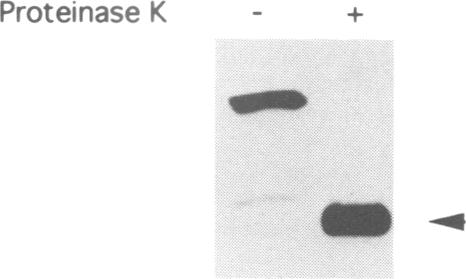
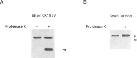
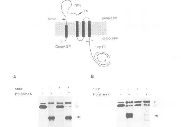
The ProW protein, located in the inner membrane of Escherichia coli, has a very unusual topology with a 100-residue-long N-terminal tail protruding into the periplasmic space. We have studied the mechanism of membrane translocation of the periplasmic tail by analysing ProW-PhoA and ProW-Lep fusion proteins, both in wild-type cells and in cells with an impaired sec machinery. Our results show that the translocation efficiency is not affected by treatments that compromise the SecA and SecY functions, but that translocation is completely blocked by dissipation of the proton motive force or by the introduction of extra positively charged residues into the N-terminal tail. This suggests that the sec machinery can act properly only on domains located on the C-terminal side of a translocation signal, and that the N-terminal tail is driven through the membrane by a mechanism that involves the proton motive force.
ProW蛋白位于大肠杆菌的内膜中,其拓扑结构非常独特,有一条100个氨基酸残基长的N端尾巴伸入周质空间。我们通过分析ProW-PhoA和ProW-Lep融合蛋白,在野生型细胞和sec机制受损的细胞中研究了周质尾巴的膜转运机制。我们的结果表明,转运效率不受损害SecA和SecY功能的处理的影响,但质子动力势的耗散或在N端尾巴中引入额外的带正电荷残基会完全阻断转运。这表明sec机制只能作用于转运信号C端一侧的结构域,并且N端尾巴是通过一种涉及质子动力势的机制被驱动穿过膜的。