Chacon E, Reece J M, Nieminen A L, Zahrebelski G, Herman B, Lemasters J J
Department of Cell Biology & Anatomy, University of North Carolina, Chapel Hill 27599.
Biophys J. 1994 Apr;66(4):942-52. doi: 10.1016/S0006-3495(94)80904-X.
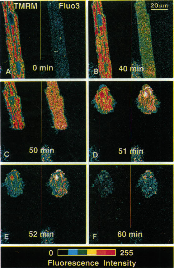
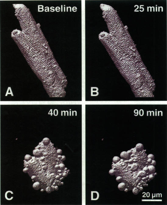
Exploiting the optical sectioning capabilities of laser scanning confocal microscopy and using parameter-specific fluorescent probes, we determined the distribution of pH, free Ca2+, electrical potential, and volume inside cultured adult rabbit cardiac myocytes during ATP depletion and reductive stress with cyanide and 2-deoxyglucose ("chemical hypoxia"). During normoxic incubations, myocytes exhibited a cytosolic pH of 7.1 and a mitochondrial pH of 8.0 (delta pH = 0.9 units). Sarcolemmal membrane potential (delta psi) was -80 mV, and mitochondrial delta psi was as high as -100 mV, yielding a mitochondrial protonmotive force (delta p) of -155 mV (delta P = delta psi - 60 delta pH). After 30 min of chemical hypoxia, mitochondrial delta pH decreased to 0.5 pH units, but mitochondrial delta psi remained essentially unchanged. By 40 min, delta pH was collapsed, and mitochondrial and cytosolic free Ca2+ began to increase. Mitochondrial and sarcolemmal delta psi remained high. as Ca2+ rose, myocytes shortened, hypercontracted, and blebbed with a 30% decrease of cell volume. After hypercontraction, extensive mitochondrial Ca2+ loading occurred. After another few minutes, mitochondrial depolarized completely and released their load of Ca2+. After many more minutes, the sarcolemmal permeability barrier broke down, and viability was lost. These studies demonstrate a sequence of subcellular ionic and electrical changes that may underlie the progression to irreversible hypoxic injury.
利用激光扫描共聚焦显微镜的光学切片能力并使用参数特异性荧光探针,我们测定了培养的成年兔心肌细胞在ATP耗竭以及用氰化物和2-脱氧葡萄糖(“化学性缺氧”)诱导的还原应激过程中细胞内pH、游离Ca2+、电势和体积的分布。在常氧孵育期间,心肌细胞的胞质pH为7.1,线粒体pH为8.0(pH差值 = 0.9单位)。肌膜膜电位(Δψ)为 -80 mV,线粒体Δψ高达 -100 mV,产生的线粒体质子动力(Δp)为 -155 mV(ΔP = Δψ - 60ΔpH)。化学性缺氧30分钟后,线粒体pH差值降至0.5个pH单位,但线粒体Δψ基本保持不变。到40分钟时,pH差值消失,线粒体和胞质游离Ca2+开始增加。线粒体和肌膜的Δψ仍然很高。随着Ca2+升高,心肌细胞缩短、过度收缩并出现泡状改变,细胞体积减少30%。过度收缩后,线粒体发生广泛的Ca2+加载。再过几分钟后,线粒体完全去极化并释放其Ca2+负荷。又过了许多分钟后,肌膜通透性屏障破坏,细胞失去活力。这些研究证明了一系列亚细胞离子和电变化,这些变化可能是不可逆性缺氧损伤进展的基础。