• 文献检索
  • 文档翻译
  • 深度研究
  • 学术资讯
  • Suppr Zotero 插件Zotero 插件
  • 邀请有礼
  • 套餐&价格
  • 历史记录
应用&插件
Suppr Zotero 插件Zotero 插件浏览器插件Mac 客户端Windows 客户端微信小程序
定价
高级版会员购买积分包购买API积分包
服务
文献检索文档翻译深度研究API 文档MCP 服务
关于我们
关于 Suppr公司介绍联系我们用户协议隐私条款
关注我们

Suppr 超能文献

核心技术专利:CN118964589B侵权必究
粤ICP备2023148730 号-1Suppr @ 2026

文献检索

告别复杂PubMed语法,用中文像聊天一样搜索,搜遍4000万医学文献。AI智能推荐,让科研检索更轻松。

立即免费搜索

文件翻译

保留排版,准确专业,支持PDF/Word/PPT等文件格式,支持 12+语言互译。

免费翻译文档

深度研究

AI帮你快速写综述,25分钟生成高质量综述,智能提取关键信息,辅助科研写作。

立即免费体验

上皮性卵巢癌的间期分子细胞遗传学分析

Interphase molecular cytogenetic analysis of epithelial ovarian carcinomas.

作者信息

Persons D L, Hartmann L C, Herath J F, Borell T J, Cliby W A, Keeney G L, Jenkins R B

机构信息

Department of Laboratory Medicine and Pathology, Mayo Clinic/Foundation, Rochester, Minnesota.

出版信息

Am J Pathol. 1993 Mar;142(3):733-41.

PMID:8096121
原文链接:https://pmc.ncbi.nlm.nih.gov/articles/PMC1886787/
Abstract

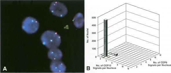
Karyotype information on ovarian carcinomas has been limited because the tumors are often difficult to culture and the resultant metaphases can have complex numerical and structural chromosomal anomalies. Fluorescent in situ hybridization is a rapid method of determining centromere copy number in metaphase cells and interphase nuclei. Fluorescent in situ hybridization was used to determine the numerical centromere complement of chromosomes X, 8, 12, and 17 and HER-2/neu gene amplification within interphase nuclei of 25 primary epithelial ovarian carcinomas. Touch preparations of the carcinomas were hybridized with two-color combinations of directly labeled alpha-satellite centromeric chromosome enumeration probes and a directly labeled HER-2/neu probe. Modal centromere copy numbers for each of the four chromosomes were used to determine numerical abnormalities relative to the flow cytometric DNA ploidy level for each tumor. Four cases were found to be normal with respect to the four chromosomes studied. In the remaining 21 cases a relative loss of chromosomes 17 (16 cases) and X (nine cases) and a relative gain of chromosomes 12 (10 cases) and 8 (nine cases) were the most common findings. In addition, the HER-2/neu gene was amplified in two of the 25 tumors. In conclusion, fluorescent in situ hybridization is an excellent method for rapid determination of numerical abnormalities and gene amplification in ovarian carcinomas.

摘要

关于卵巢癌的核型信息一直有限,因为这些肿瘤通常难以培养,并且所得到的中期细胞可能具有复杂的染色体数目和结构异常。荧光原位杂交是一种在中期细胞和间期核中确定着丝粒拷贝数的快速方法。荧光原位杂交被用于确定25例原发性上皮性卵巢癌间期核内染色体X、8、12和染色体17的着丝粒数目以及HER-2/neu基因扩增情况。将癌组织的触片与直接标记的α-卫星着丝粒染色体计数探针和直接标记的HER-2/neu探针的双色组合进行杂交。使用这四条染色体各自的着丝粒拷贝数众数来确定相对于每个肿瘤的流式细胞术DNA倍体水平的数目异常情况。在所研究的四条染色体方面,发现有4例正常。在其余21例中,最常见的发现是染色体17(16例)和X(9例)相对缺失,以及染色体12(10例)和8(9例)相对增加。此外,25例肿瘤中有2例HER-2/neu基因发生扩增。总之,荧光原位杂交是快速确定卵巢癌数目异常和基因扩增的一种出色方法。

https://cdn.ncbi.nlm.nih.gov/pmc/blobs/4874/1886787/15e99a7784c8/amjpathol00075-0081-a.jpg
https://cdn.ncbi.nlm.nih.gov/pmc/blobs/4874/1886787/ff403005d3f7/amjpathol00075-0079-a.jpg
https://cdn.ncbi.nlm.nih.gov/pmc/blobs/4874/1886787/68864fa3161e/amjpathol00075-0079-b.jpg
https://cdn.ncbi.nlm.nih.gov/pmc/blobs/4874/1886787/15e99a7784c8/amjpathol00075-0081-a.jpg
https://cdn.ncbi.nlm.nih.gov/pmc/blobs/4874/1886787/ff403005d3f7/amjpathol00075-0079-a.jpg
https://cdn.ncbi.nlm.nih.gov/pmc/blobs/4874/1886787/68864fa3161e/amjpathol00075-0079-b.jpg
https://cdn.ncbi.nlm.nih.gov/pmc/blobs/4874/1886787/15e99a7784c8/amjpathol00075-0081-a.jpg

相似文献

1
Interphase molecular cytogenetic analysis of epithelial ovarian carcinomas.上皮性卵巢癌的间期分子细胞遗传学分析
Am J Pathol. 1993 Mar;142(3):733-41.
2
c-myc and chromosome 8 centromere studies of ovarian cancer by interphase FISH.应用间期荧光原位杂交技术对卵巢癌进行c-myc和8号染色体着丝粒研究。
Exp Mol Pathol. 1999 Jun;66(2):140-8. doi: 10.1006/exmp.1999.2259.
3
Interphase cytogenetic analysis of mucinous ovarian neoplasms.黏液性卵巢肿瘤的间期细胞遗传学分析
Lab Invest. 1997 May;76(5):661-70.
4
Detection of numerical chromosome anomalies in interphase cells of ovarian carcinomas using fluorescence in situ hybridization.利用荧光原位杂交技术检测卵巢癌间期细胞中的染色体数目异常
Genes Chromosomes Cancer. 1996 Jun;16(2):120-9. doi: 10.1002/(SICI)1098-2264(199606)16:2<120::AID-GCC6>3.0.CO;2-1.
5
Interphase cytogenetic analysis of serous ovarian tumors of low malignant potential: comparison with serous cystadenomas and invasive serous carcinomas.低恶性潜能浆液性卵巢肿瘤的间期细胞遗传学分析:与浆液性囊腺瘤及浸润性浆液性癌的比较
Lab Invest. 1996 Oct;75(4):473-85.
6
HER-2/neu oncogene amplification in stage I and stage III ovarian papillary serous carcinoma.HER-2/neu原癌基因在Ⅰ期和Ⅲ期卵巢乳头状浆液性癌中的扩增
Exp Mol Pathol. 1999 Jun;66(2):163-9. doi: 10.1006/exmp.1999.2255.
7
Interphase cytogenetic studies of human hepatocellular carcinomas by fluorescent in situ hybridization.通过荧光原位杂交技术对人肝细胞癌进行间期细胞遗传学研究。
Hepatology. 1996 Mar;23(3):429-35. doi: 10.1002/hep.510230306.
8
Flow cytometric quantification of human chromosome specific repetitive DNA sequences by single and bicolor fluorescent in situ hybridization to lymphocyte interphase nuclei.通过对淋巴细胞间期核进行单色和双色荧光原位杂交,采用流式细胞术对人类染色体特异性重复DNA序列进行定量分析。
Cytometry. 1990;11(1):153-64. doi: 10.1002/cyto.990110118.
9
Her-2/neu expression and amplification in early stage ovarian surface epithelial neoplasms.Her-2/neu在早期卵巢表面上皮性肿瘤中的表达及扩增
Gynecol Oncol. 2004 Dec;95(3):570-5. doi: 10.1016/j.ygyno.2004.08.043.
10
Interphase cytogenetics of prostatic tumor progression: specific chromosomal abnormalities are involved in metastasis to the bone.前列腺肿瘤进展的间期细胞遗传学:特定染色体异常与骨转移有关。
Lab Invest. 1997 Nov;77(5):437-48.

引用本文的文献

1
Consistent numerical chromosome aberrations in thecofibromas of the ovary.卵巢纤维瘤中一致的染色体数目异常。
Virchows Arch. 2008 Mar;452(3):269-76. doi: 10.1007/s00428-007-0561-x.
2
Molecular cytogenetic characterization of tenosynovial giant cell tumors.腱鞘巨细胞瘤的分子细胞遗传学特征
Neoplasia. 2004 Sep-Oct;6(5):578-83. doi: 10.1593/neo.04202.
3
Evaluation of the tyrosine kinase domain of the Met proto-oncogene in sporadic ovarian carcinomas*.散发性卵巢癌中Met原癌基因酪氨酸激酶结构域的评估*

本文引用的文献

1
Karyotype analysis of four solid gynecologic tumors.四种妇科实体肿瘤的核型分析
Gynecol Oncol. 1982 Dec;14(3):324-38. doi: 10.1016/0090-8258(82)90107-x.
2
Method for analysis of cellular DNA content of paraffin-embedded pathological material using flow cytometry.使用流式细胞术分析石蜡包埋病理材料细胞DNA含量的方法。
J Histochem Cytochem. 1983 Nov;31(11):1333-5. doi: 10.1177/31.11.6619538.
3
A detergent-trypsin method for the preparation of nuclei for flow cytometric DNA analysis.一种用于制备细胞核以进行流式细胞术DNA分析的去污剂-胰蛋白酶法。
Pathol Oncol Res. 1999;5(3):187-91. doi: 10.1053/paor.1999.0219.
4
Amplification of the c-erbB-2 (HER-2/neu) gene in gastric cancer cells. Detection by fluorescence in situ hybridization.胃癌细胞中c-erbB-2(HER-2/neu)基因的扩增。荧光原位杂交检测
Am J Pathol. 1997 Sep;151(3):761-8.
5
Interphase cytogenetics of prostate cancer: fluorescence in situ hybridisation (FISH) analysis of Japanese cases.前列腺癌的间期细胞遗传学:日本病例的荧光原位杂交(FISH)分析
Br J Cancer. 1996 Dec;74(11):1699-704. doi: 10.1038/bjc.1996.617.
6
Fluorescent in situ hybridization assessment of chromosome 8 copy number in breast cancer.乳腺癌中8号染色体拷贝数的荧光原位杂交评估
Breast Cancer Res Treat. 1996;38(2):201-8. doi: 10.1007/BF01806674.
7
Interphase in situ hybridization to disaggregated and intact tissue specimens of prostatic adenocarcinomas.前列腺腺癌解离和完整组织标本的间期原位杂交
Histochem Cell Biol. 1995 Dec;104(6):479-86. doi: 10.1007/BF01464339.
8
Origins of heterogeneous ovarian carcinomas. A molecular cytogenetic analysis of histologically benign, low malignant potential, and fully malignant components.异质性卵巢癌的起源。对组织学良性、低恶性潜能和完全恶性成分的分子细胞遗传学分析。
Am J Pathol. 1996 Aug;149(2):511-20.
9
The genetic analysis of ovarian cancer.卵巢癌的基因分析
Br J Cancer. 1995 Sep;72(3):521-7. doi: 10.1038/bjc.1995.367.
10
Novel fluorescence in situ hybridization approaches in solid tumors. Characterization of frozen specimens, touch preparations, and cytological preparations.实体瘤中的新型荧光原位杂交方法。冷冻标本、涂片标本和细胞学标本的特征分析。
Am J Pathol. 1995 Oct;147(4):896-904.
Cytometry. 1983 Mar;3(5):323-7. doi: 10.1002/cyto.990030503.
4
Specificity of proto-oncogene amplification in human malignant diseases.人类恶性疾病中原癌基因扩增的特异性
Mol Biol Med. 1987 Aug;4(4):213-27.
5
Cytogenetic analysis of four human ovarian carcinoma cell lines.四种人卵巢癌细胞系的细胞遗传学分析。
Cancer Genet Cytogenet. 1987 Jun;26(2):339-49. doi: 10.1016/0165-4608(87)90068-9.
6
Cytogenetic findings in cell lines derived from four ovarian carcinomas.源自四种卵巢癌的细胞系的细胞遗传学发现。
Cancer Genet Cytogenet. 1987 Feb;24(2):231-42. doi: 10.1016/0165-4608(87)90104-x.
7
Cytogenetic analysis using quantitative, high-sensitivity, fluorescence hybridization.使用定量、高灵敏度荧光杂交技术的细胞遗传学分析。
Proc Natl Acad Sci U S A. 1986 May;83(9):2934-8. doi: 10.1073/pnas.83.9.2934.
8
Abnormal chromosomes including small metacentrics in 14 ovarian cancers.14例卵巢癌中存在包括小中着丝粒染色体在内的异常染色体。
Cancer Genet Cytogenet. 1987 Jun;26(2):355-61. doi: 10.1016/0165-4608(87)90070-7.
9
Uterine leiomyoma cytogenetics. I. Rearrangements of chromosome 12.子宫平滑肌瘤细胞遗传学。I. 12号染色体重排。
Cancer Genet Cytogenet. 1989 Jan;37(1):49-54. doi: 10.1016/0165-4608(89)90073-3.
10
Consistent occurrence of a 19p+ marker chromosome and loss of 11p material in ovarian seropapillary cystadenocarcinomas.卵巢浆液性乳头状囊腺癌中19号染色体短臂增加标记染色体及11号染色体短臂物质缺失的一致性出现。
Genes Chromosomes Cancer. 1989 Nov;1(2):167-71. doi: 10.1002/gcc.2870010210.