Keitel T, Simon O, Borriss R, Heinemann U
Institut für Kristallographie, Freie Universität, Berlin, Federal Republic of Germany.
Proc Natl Acad Sci U S A. 1993 Jun 1;90(11):5287-91. doi: 10.1073/pnas.90.11.5287.
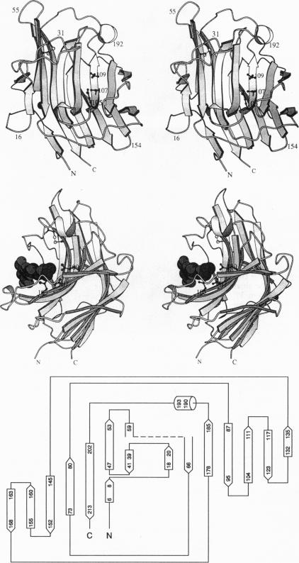
The three-dimensional structure of the hybrid Bacillus 1,3-1,4-beta-glucanase (beta-glucanase; 1,3-1,4-beta-D-glucan 4-glucanohydrolase, lichenase, EC 3.2.1.73) designated H(A16-M) was determined by x-ray crystallography at a resolution of 2.0 A and refined to an R value of 16.4% using stereochemical restraints. The protein molecule consists mainly of two seven-stranded antiparallel beta-pleated sheets arranged atop each other to form a compact, sandwich-like structure. A channel crossing one side of the protein molecule accommodates an inhibitor, 3,4-epoxybutyl beta-D-cellobioside, which binds covalently to the side chain of Glu-105, as seen in a crystal structure analysis at 2.8-A resolution of the protein-inhibitor complex (R = 16.8%). That Glu-105 may be indispensible for enzyme catalysis by H(A16-M) is suggested by site-directed mutagenesis of this residue, which inevitably leads to an inactive enzyme.
通过X射线晶体学在2.0埃的分辨率下确定了命名为H(A16 - M)的杂交芽孢杆菌1,3 - 1,4-β-葡聚糖酶(β-葡聚糖酶;1,3 - 1,4-β-D-葡聚糖4-葡聚糖水解酶,地衣多糖酶,EC 3.2.1.73)的三维结构,并使用立体化学限制将其精修至R值为16.4%。蛋白质分子主要由两个七股反平行β折叠片层相互堆叠形成紧密的三明治状结构组成。蛋白质分子一侧的一个通道容纳一种抑制剂3,4-环氧丁基β-D-纤维二糖苷,如在蛋白质-抑制剂复合物2.8埃分辨率的晶体结构分析(R = 16.8%)中所见,它与Glu-105的侧链共价结合。对该残基进行定点诱变不可避免地导致无活性的酶,这表明Glu-105对于H(A16 - M)的酶催化可能是必不可少的。