Wolfson M, Lev M, Avinoah I, Malik Z, Löchelt M, Flügel R M, Dombrovski A, Aboud M
Department of Microbiology and Immunology, Faculty of Health Sciences, Ben Gurion University of the Negev, Beer Sheva, Israel.
J Virol. 1994 Jul;68(7):4695-9. doi: 10.1128/JVI.68.7.4695-4699.1994.
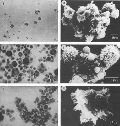
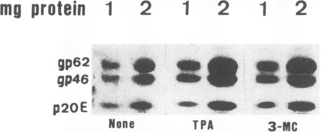
Treatment of human T-cell leukemia virus type I (HTLV-I)- and HTLV-II-infected T-cell lines with 12-O-tetradecanoylphorbol-13-acetate (TPA) stimulated virus release. However, this stimulation was mainly detected at 42 to 48 h of treatment, whereas later virus release declined rapidly. During the first 48 h, TPA had no effect on cell growth, but later, the number of viable cells was profoundly lower in the TPA-treated than in the untreated cultures. This shift in virus release and cell number resulted from self-fusion of a large proportion of the virus-producing cells, which seemed to consequently enter into a dying process. This fusion, which resulted in syncytium formation, was strongly inhibited by anti-HTLV-I env monoclonal antibodies. Furthermore, no self-fusion was detected in three different uninfected T-cell lines similarly treated with TPA. On the other hand, stimulation of virus production by 3-methylcholanthrene (3-MC) treatment failed to induce self-fusion in the infected cells. Moreover, no syncytium was detected when these 3-MC-treated infected cells were cocultured with any of the TPA-treated uninfected cells. The effects of TPA on virus production and syncytium formation were both abolished by three different protein kinase C inhibitors. Taken together, these data suggest that the self-fusion observed in these experiments required both enhanced virus production and protein kinase C-phosphorylated viral or/and virally induced cellular component(s).
用12 - O - 十四烷酰佛波醇 - 13 - 乙酸酯(TPA)处理人I型嗜T细胞病毒(HTLV - I)和HTLV - II感染的T细胞系可刺激病毒释放。然而,这种刺激主要在处理42至48小时时检测到,而随后病毒释放迅速下降。在最初的48小时内,TPA对细胞生长没有影响,但之后,TPA处理的培养物中活细胞数量比未处理的培养物中显著减少。病毒释放和细胞数量的这种变化是由于大部分产生病毒的细胞发生自融合,这似乎导致细胞进入死亡过程。这种导致多核巨细胞形成的融合受到抗HTLV - I env单克隆抗体的强烈抑制。此外,在用TPA进行类似处理的三种不同的未感染T细胞系中未检测到自融合。另一方面,用3 - 甲基胆蒽(3 - MC)处理刺激病毒产生未能在感染细胞中诱导自融合。此外,当这些用3 - MC处理的感染细胞与任何用TPA处理的未感染细胞共培养时,未检测到多核巨细胞。三种不同的蛋白激酶C抑制剂均消除了TPA对病毒产生和多核巨细胞形成的影响。综上所述,这些数据表明,在这些实验中观察到的自融合既需要增强的病毒产生,也需要蛋白激酶C磷酸化的病毒或/和病毒诱导的细胞成分。