Hollander A P, Pidoux I, Reiner A, Rorabeck C, Bourne R, Poole A R
Joint Diseases Laboratory, Shriners Hospital for Crippled Children, Montreal, Quebec, Canada.
J Clin Invest. 1995 Dec;96(6):2859-69. doi: 10.1172/JCI118357.
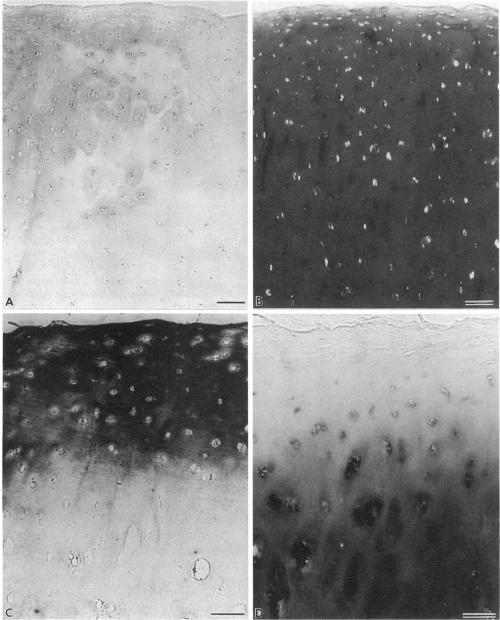
Enhanced denaturation of type II collagen fibrils in femoral condylar cartilage in osteoarthritis (OA) has recently been quantitated immunochemically (Hollander, A.P., T.F. Heathfield, C. Webber, Y. Iwata, R. Bourne, C. Rorabeck, and A.R. Poole. 1994. J. Clin. Invest. 93:1722-1732). Using the same antibody that only reacts with denatured type II collagen, we investigated with immunoperoxidase histochemistry (results were graded for analysis) the sites of the denaturation (loss of triple helix) of this molecule in human aging (at autopsy, n= 11) and progressively degenerate (by Mankin grade [MG]) OA (at arthroplasty, n= 51) knee condylar cartilages. Up to 41 yr, most aging cartilages (3 of 4) (MG 0-4) showed very little denaturation. In most older cartilages, (4 of 7) (MG 2-4), staining was observed in the superficial and mid zones. This pattern of collagen II denaturation was also seen in all OA specimens with increased staining extending to the deep zone with increasing MG. Collagen II staining correlated directly both with MG and collagen II denaturation measured by immunoassay. Cartilage fibrillation occurred in OA cartilages with increased penetration of the staining for collagen II denaturation into the mid and deep zones and where denaturation was more pronounced by immunoassay. Thus in both aging and OA the first damage to type II collagen occurs in the superficial and upper mid zone (low MG) extending to the lower mid and deep zones with increasing degeneration (increasing MG). Initial damage is always seen around chondrocytes implicating them in the denaturation of type II collagen.
最近,通过免疫化学方法对骨关节炎(OA)患者股骨髁软骨中II型胶原纤维的变性增强进行了定量研究(霍兰德,A.P.,T.F.希思菲尔德,C.韦伯,Y.岩田,R.伯恩,C.罗勒贝克,和A.R.普尔。1994年。《临床研究杂志》93:1722 - 1732)。我们使用仅与变性II型胶原反应的相同抗体,通过免疫过氧化物酶组织化学方法(对结果进行分级分析)研究了该分子在人类衰老(尸检,n = 11)和逐渐退变(按曼金分级[MG])的OA(关节置换术时,n = 51)膝关节髁软骨中的变性(三螺旋结构丧失)部位。在41岁之前,大多数衰老软骨(4个中的3个)(MG 0 - 4)显示出很少的变性。在大多数老年软骨(7个中的4个)(MG 2 - 4)中,在表层和中层区域观察到染色。在所有OA标本中也观察到这种II型胶原变性模式,随着MG增加,染色增加并延伸至深层区域。II型胶原染色与MG以及通过免疫测定法测量的II型胶原变性直接相关。在OA软骨中出现了软骨纤维化,II型胶原变性染色向中层和深层区域的渗透增加,并且通过免疫测定法变性更明显。因此,在衰老和OA中,II型胶原的最初损伤都发生在表层和上中层区域(低MG),随着退变加剧(MG增加)延伸至中下层和深层区域。最初的损伤总是在软骨细胞周围观察到,这表明软骨细胞与II型胶原的变性有关。