Dodge G R, Poole A R
Shriners Hospital for Crippled Children, Department of Surgery, McGill University, Montreal, Quebec, Canada.
J Clin Invest. 1989 Feb;83(2):647-61. doi: 10.1172/JCI113929.
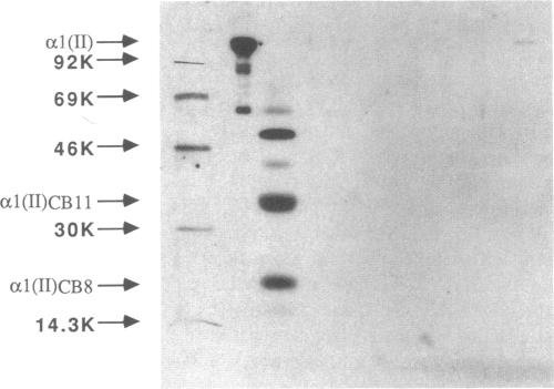
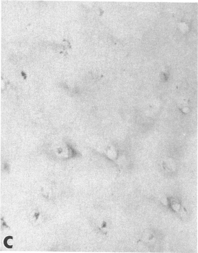
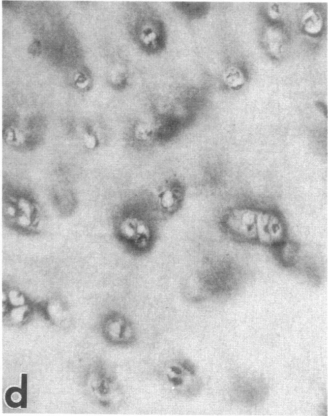
Articular cartilage destruction and loss of function in arthritic diseases involves proteolytic degradation of the connective tissue matrix. We have investigated the degradation of cartilage collagen by developing immunochemical methods that permit the identification and analysis of type II collagen degradation in situ. Previously, a technique to specifically identify type II collagen degradation in situ in articular cartilage did not exist. These methods utilize a polyclonal antiserum (R181) that specifically reacts with unwound alpha-chains and CNBr-derived peptides, alpha 1(II)CB11 and alpha 1(II)CB8, of human and bovine type II collagens. The experimental approach is based on the fact that when fibrillar collagens are cleaved the helical collagen molecule unwinds, exposing hidden epitopes. Here we demonstrate the use of R181 in studying type II collagen degradation in bovine articular cartilage that has been cultured with or without IL-1 and in human normal, rheumatoid, and osteoarthritic articular cartilages. Compared to cartilages either freshly isolated or cultured without IL-1, bovine cartilage cultured with IL-1 for 3-5 d showed an increase in both pericellular and intercellular immunohistochemical staining. Extracts of these cartilages contained type II collagen alpha chains that were increased in amount after culture with IL-1 for 11 d. In addition, culture with IL-1 resulted in the appearance of alpha chain fragments of lower molecular weight. All human arthritic tissues examined showed areas of pronounced pericellular and territorial staining for collagen degradation as compared with non-diseased tissues, indicating that chondrocytes are responsible in part for this degradation as compared with non-diseased tissues. In most cases rheumatoid cartilage was stained most intensely at the articular surface and in the deep and mid-zones, whereas osteoarthritic cartilage usually stained more in the superficial and mid-zones, but less intensely. Distinct patterns of sites of collagen degradation reflect differences in collagen destruction in these diseases, suggesting possible different sources of chondrocyte activation. These experiments demonstrate the application of immunological methods to detect collagen degradation and demonstrate an increase of collagen degradation in human arthritides and in IL-1-treated viable bovine cartilage.
关节炎性疾病中关节软骨的破坏和功能丧失涉及结缔组织基质的蛋白水解降解。我们通过开发免疫化学方法来研究软骨胶原蛋白的降解,这些方法能够在原位鉴定和分析II型胶原蛋白的降解。以前,不存在特异性鉴定关节软骨中II型胶原蛋白原位降解的技术。这些方法利用了一种多克隆抗血清(R181),它能与人及牛II型胶原蛋白的解旋α链以及CNBr衍生肽α1(II)CB11和α1(II)CB8特异性反应。实验方法基于这样一个事实,即当纤维状胶原蛋白被切割时,螺旋状的胶原蛋白分子会解旋,暴露出隐藏的表位。在这里,我们展示了R181在研究牛关节软骨中II型胶原蛋白降解的应用,这些软骨在有或无白细胞介素-1(IL-1)的条件下进行培养,以及在人类正常、类风湿性和骨关节炎性关节软骨中的应用。与新鲜分离或在无IL-1条件下培养的软骨相比,用IL-1培养3 - 5天的牛软骨在细胞周围和细胞间的免疫组织化学染色均增加。这些软骨的提取物含有II型胶原蛋白α链,在用IL-1培养11天后其数量增加。此外,用IL-1培养导致出现分子量较低的α链片段。与非患病组织相比,所有检查的人类关节炎组织均显示出明显的细胞周围和区域胶原降解染色区域,表明与非患病组织相比,软骨细胞在这种降解中起部分作用。在大多数情况下,类风湿性软骨在关节表面以及深部和中间区域染色最强烈,而骨关节炎性软骨通常在浅表和中间区域染色较多,但强度较低。胶原蛋白降解部位的不同模式反映了这些疾病中胶原蛋白破坏的差异,提示软骨细胞激活可能存在不同来源。这些实验证明了免疫方法在检测胶原蛋白降解中的应用,并证明了人类关节炎以及IL-1处理的活牛软骨中胶原蛋白降解增加。