Pidoux A L, LeDizet M, Cande W Z
Department of Molecular and Cell Biology, University of California, Berkeley 94720-3200, USA.
Mol Biol Cell. 1996 Oct;7(10):1639-55. doi: 10.1091/mbc.7.10.1639.
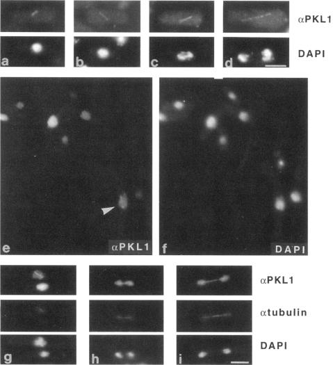
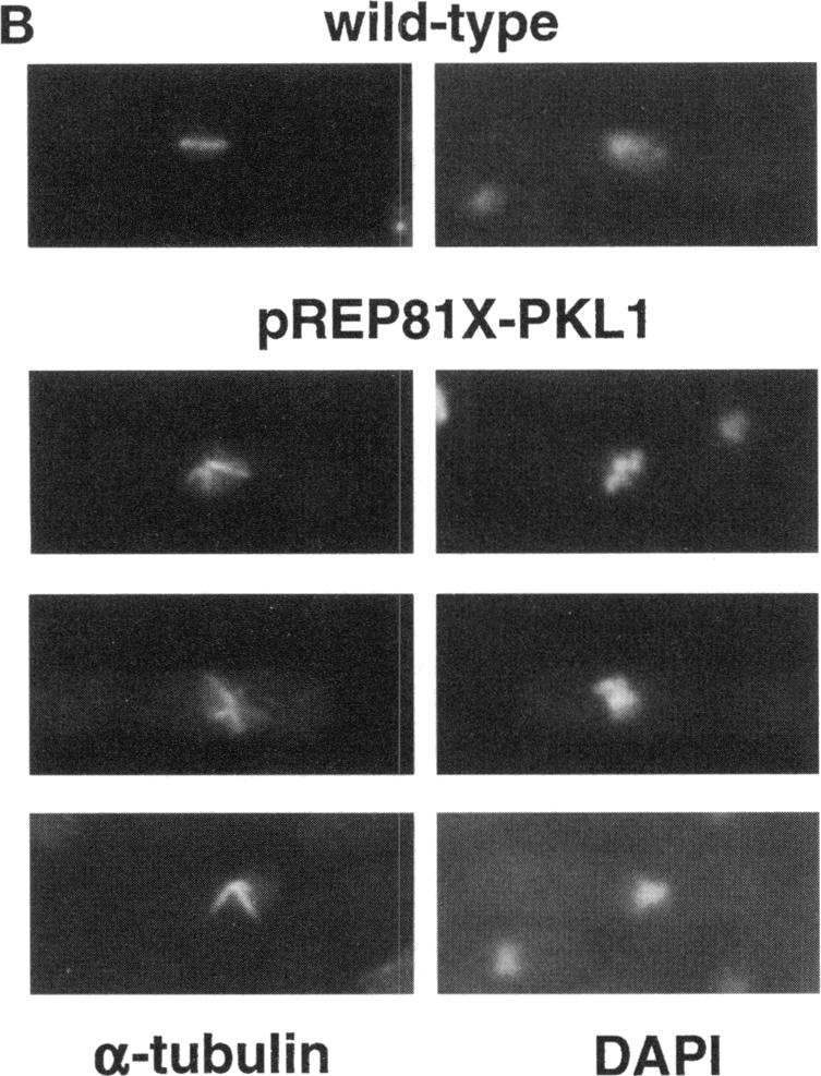
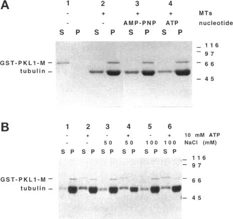
We have used anti-peptide antibodies raised against highly conserved regions of the kinesin motor domain to identify kinesin-related proteins in the fission yeast Schizosaccharomyces pombe. Here we report the identification of a new kinesin-related protein, which we have named pkl1. Sequence homology and domain organization place pkl1 in the Kar3/ncd subfamily of kinesin-related proteins. Bacterially expressed pkl1 fusion proteins display microtubule-stimulated ATPase activity, nucleotide-sensitive binding, and bundling of microtubules. Immunofluorescence studies with affinity-purified antibodies indicate that the pkl1 protein localizes to the nucleus and the mitotic spindle. Pkl1 null mutants are viable but have increased sensitivity to microtubule-disrupting drugs. Disruption of pkl1+ suppresses mutations in another kinesin-related protein, cut7, which is known to act in the spindle. Overexpression of pkl1 to very high levels causes a similar phenotype to that seen in cut7 mutants: V-shaped and star-shaped microtubule structures are observed, which we interpret to be spindles with unseparated spindle poles. These observations suggest that pkl1 and cut7 provide opposing forces in the spindle. We propose that pkl1 functions as a microtubule-dependent motor that is involved in microtubule organization in the mitotic spindle.
我们利用针对驱动蛋白运动结构域高度保守区域产生的抗肽抗体,在裂殖酵母粟酒裂殖酵母中鉴定与驱动蛋白相关的蛋白质。在此,我们报告鉴定出一种新的与驱动蛋白相关的蛋白质,我们将其命名为pkl1。序列同源性和结构域组织将pkl1归入驱动蛋白相关蛋白质的Kar3/ncd亚家族。细菌表达的pkl1融合蛋白表现出微管刺激的ATP酶活性、核苷酸敏感结合以及微管成束。用亲和纯化抗体进行的免疫荧光研究表明,pkl1蛋白定位于细胞核和有丝分裂纺锤体。Pkl1基因敲除突变体是可存活的,但对破坏微管的药物敏感性增加。破坏pkl1+可抑制另一种已知在纺锤体中起作用的与驱动蛋白相关的蛋白质cut7中的突变。将pkl1过度表达至非常高的水平会导致与cut7突变体中所见相似的表型:观察到V形和星形微管结构,我们将其解释为纺锤体极未分离的纺锤体。这些观察结果表明,pkl1和cut7在纺锤体中提供相反的力。我们提出,pkl1作为一种微管依赖性马达,参与有丝分裂纺锤体中的微管组织。