Norose K, Yano A
Department of Ophthalmology, Shinshu University School of Medicine, Matsumoto, Japan.
Br J Ophthalmol. 1996 Nov;80(11):1002-8. doi: 10.1136/bjo.80.11.1002.
AIMS/BACKGROUND: To determine the functional properties and cytokine production profiles of melanoma specific cytotoxic T lymphocytes (CTLs) induced from peripheral blood leucocytes of two patients with Vogt-Koyanagi-Harada disease (VKH).
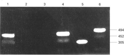
Melanoma specific CTL lines were established by long term coculture with a human melanoma cell line (P-36). Cytotoxic activity against P-36 was measured by 51Cr release. The involvement of human leucocyte antigen (HLA) class I or class II molecules in the cytotoxicity of the CTL lines against P-36 was analysed using anti-HLA class I or anti-HLA class II monoclonal antibody (MAb). Surface molecules of CTL lines were analysed by flow cytometry using MAbs specific for CD4, CD8, CD16, CD25, CD56, HLA-DR, T cell antigen receptor (TCR) alpha beta and TCR gamma delta. Cytokine production and soluble interleukin 2 receptor (sIL-2R) secretion were determined by enzyme linked immunosorbent assays. mRNAs of cytokines were analysed using reverse transcription polymerase chain reaction (RT-PCR).
CTLs showed strong cytotoxic activity against P-36. The CTL activity of the cell lines against P-36 was inhibited by the anti-HLA-DR MAb, whereas the MAb specific for monomorphic determinants of HLA-A, B, and C failed to block lytic activity. Flow cytometry identified the following surface molecules: CD4+, CD8-, CD16-, CD25+, CD56-, HLA-DR+, TCR alpha beta +, and TCR gamma delta-. CTLs constitutively produced a high level of IL-6. IL-6 production and sIL-2R secretion of CTLs were enhanced when CTLs were stimulated with P-36. CTLs also produced high levels of interferon gamma (IFN-gamma) and IL-2, but not IL-4. mRNAs of IL-2 and IFN-gamma were detected by RT-PCR in the CTLs.
Melanoma specific HLADR restricted T helper 1 (Th1) CTLs may play a role in the immunopathogenesis of VKH.
目的/背景:确定从两名葡萄膜大脑炎(VKH)患者外周血白细胞诱导产生的黑色素瘤特异性细胞毒性T淋巴细胞(CTL)的功能特性和细胞因子产生谱。
通过与人黑色素瘤细胞系(P-36)长期共培养建立黑色素瘤特异性CTL系。采用51Cr释放法测定对P-36的细胞毒性活性。使用抗I类或抗II类人白细胞抗原(HLA)单克隆抗体(MAb)分析HLA I类或II类分子在CTL系对P-36细胞毒性中的作用。使用针对CD4、CD8、CD16、CD25、CD56、HLA-DR、T细胞抗原受体(TCR)αβ和TCRγδ的MAb通过流式细胞术分析CTL系的表面分子。通过酶联免疫吸附测定法测定细胞因子产生和可溶性白细胞介素2受体(sIL-2R)分泌。使用逆转录聚合酶链反应(RT-PCR)分析细胞因子的mRNA。
CTL对P-36表现出强烈的细胞毒性活性。抗HLA-DR MAb可抑制细胞系对P-36的CTL活性,而针对HLA-A、B和C单态性决定簇的MAb未能阻断裂解活性。流式细胞术鉴定出以下表面分子:CD4+、CD8-、CD16-、CD25+、CD56-、HLA-DR+、TCRαβ+和TCRγδ-。CTL组成性地产生高水平的IL-6。当用P-36刺激CTL时,CTL的IL-6产生和sIL-2R分泌增强。CTL还产生高水平的干扰素γ(IFN-γ)和IL-2,但不产生IL-4。通过RT-PCR在CTL中检测到IL-2和IFN-γ的mRNA。
黑色素瘤特异性HLA-DR限制性T辅助1(Th1)CTL可能在VKH的免疫发病机制中起作用。