Esser A F, Bartholomew R M, Jensen F C, Müller-Eberhard H J
Proc Natl Acad Sci U S A. 1979 Nov;76(11):5843-7. doi: 10.1073/pnas.76.11.5843.
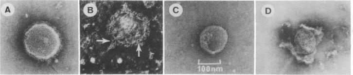
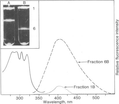
We have compared the effects of the complement membrane attack complex (MAC), nystatin, and melittin on the envelope of murine leukemia viruses to determine if channel formation alone is sufficient to cause membranolysis. Nystatin is a channel former and mellitin is not, although both are hemolytic. Whereas MAC and melittin disintegrated the viral membrane, nystatin had no effect on morphology, integrity, and infectivity of the virus. Incorporation of the antibiotic into the viral membranes was demonstrated by measurements of the characteristic fluorescence of nystatin in membranes and the dose-dependent increase in viral density after uptake of the antibiotic. The density of nystatin was measured to be 1.26-1.27 g/cm3. Proof for the formation of functional nystatin channels was obtained by light scattering measurements. Exposure of untreated virus to hypotonic conditions increased viral light scattering because of osmotic swelling but otherwise had no effect on the integrity of the virus. Nystatin channel formation abolished the light scattering change, showing that the antibiotic had impaired the viral permeability barrier. We interpret these results to indicate that virolysis by MAC is not caused by channel formation and, conversely, in the absence of colloid-osmotic effects, channel formation by itself is not sufficient to disassemble a viral membrane.
我们比较了补体膜攻击复合物(MAC)、制霉菌素和蜂毒肽对鼠白血病病毒包膜的影响,以确定仅通道形成是否足以引起膜溶解。制霉菌素是一种通道形成剂,而蜂毒肽不是,尽管两者都具有溶血作用。MAC和蜂毒肽会使病毒膜解体,而制霉菌素对病毒的形态、完整性和感染性没有影响。通过测量膜中制霉菌素的特征荧光以及摄取抗生素后病毒密度的剂量依赖性增加,证明了抗生素掺入病毒膜中。测得制霉菌素的密度为1.26 - 1.27 g/cm³。通过光散射测量获得了功能性制霉菌素通道形成的证据。未处理的病毒暴露于低渗条件下会由于渗透膨胀而增加病毒光散射,但对病毒的完整性没有其他影响。制霉菌素通道的形成消除了光散射变化,表明抗生素破坏了病毒的渗透屏障。我们对这些结果的解释是,MAC引起的病毒溶解不是由通道形成导致的,相反,在没有胶体渗透效应的情况下,通道形成本身不足以拆解病毒膜。