Rodgers F G
J Clin Pathol. 1979 Dec;32(12):1195-202. doi: 10.1136/jcp.32.12.1195.
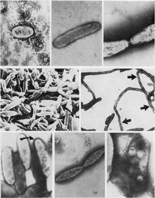
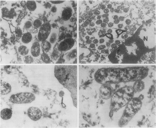
Eleven lung samples positive for Legionnaires' disease, 12 strains of Legionella pneumophila cultured on various bacteriological media, and one strain growth in the yolk sac of fertile hens' eggs were examined by negative staining, thin sectioning, and scanning electron microscopy. All organisms studied were ultrastructurally similar irrespective of strain, source, or method of cultivation, presenting mainly as short rods, 0.6 x 1.5 micrometer, with tapered ends, though long forms and filaments were also evident. In this they resembled typical Gram-negative organisms. Division was by non-septate binary fission, and the cell wall was composed of two triple-unit membranes with morphological evidence of a peptidoglycan layer. The bacterial cytoplasm was rich in ribosomes and nuclear elements and often contained vacuoles. No acid polysaccharides or bacterial appendages were detected surrounding the organisms. In lung tissue and yolk sac membranes, the organisms replicated within the cytoplasm of infected cells and in the intercellular spaces and were specifically identified in thin sections by immunoferritin techniques.