Ownby C L, Cameron D, Tu A T
Am J Pathol. 1976 Oct;85(1):149-66.
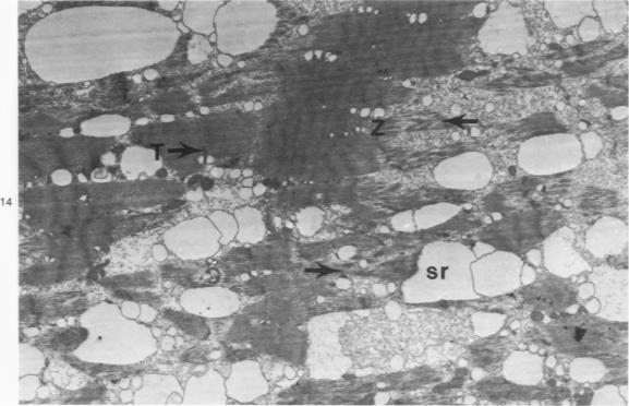
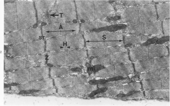
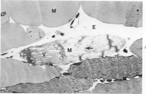
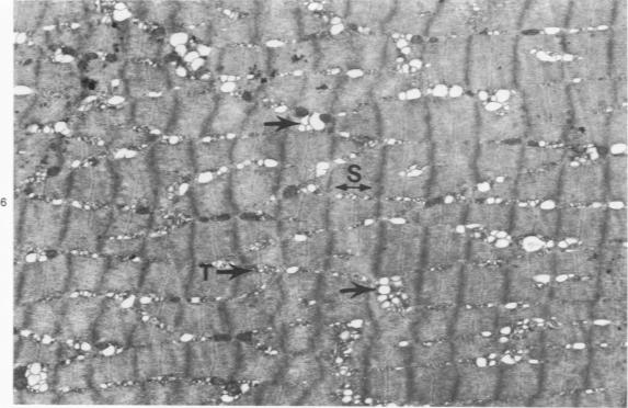
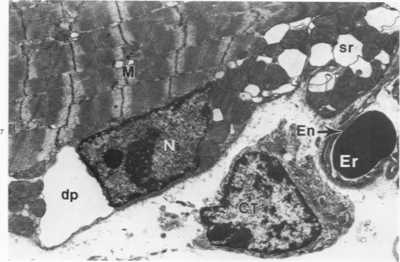
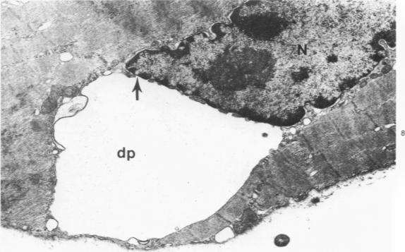
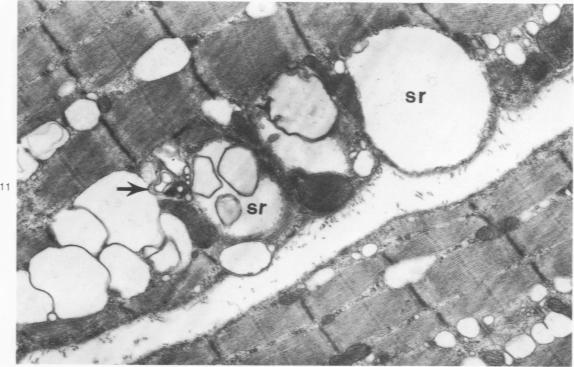
The pathogenesis of myonecrosis induced by a purified component of rattlesnake (Crotalus viridis viridis) venom was studied at the light and electron microscopic levels. Crude venom was fractionated by gel filtration (Sephadex G-50) followed by cation exchange chromatography (Sephadex C-25). Electrophoretic homogeneity of the isolated myotoxin (Fraction II from C-25 column) was demonstrated in isoelectric focusing and disc gel polyacrylamide gel electrophoresis. White mice were injected intramuscularly with 1.5 mug/g of the purified protein in 0.1 ml of physiologic saline. Light microscopic examination of injected muscle revealed a series of degenerative events including partial vacuolation of muscle cells at 6, 12, and 24 hours and complete vacuolation and loss of striations at 48 and 72 hours. Hemorrhage was not observed. At the electron microscopic level the perinuclear space and sarcoplasmic reticulum were dilated in all samples. By 48 and 72 hours the myofibrils lacked striations and the sarcomeres were disorganized. Plasma membranes and T tubules remained intact in all samples. These results correlated well with the myonecrosis induced by crude Crotalus viridis viridis venom except for several important aspects. The pure component altered skeletal muscle cells specifically, with the sarcoplasmic reticulum being the primary site of action.
在光学显微镜和电子显微镜水平上研究了响尾蛇(草原响尾蛇)毒液的一种纯化成分诱导的肌坏死的发病机制。粗毒液先通过凝胶过滤(葡聚糖G - 50)进行分级分离,然后进行阳离子交换色谱(葡聚糖C - 25)分离。在等电聚焦和圆盘凝胶聚丙烯酰胺凝胶电泳中证明了分离出的肌毒素(C - 25柱的组分II)在电泳上的均一性。将1.5微克/克的纯化蛋白溶解于0.1毫升生理盐水中,给小白鼠进行肌肉注射。对注射部位肌肉的光学显微镜检查显示出一系列退行性变化,包括在6、12和24小时时肌肉细胞出现部分空泡化,在48和72小时时出现完全空泡化且横纹消失。未观察到出血现象。在电子显微镜水平上,所有样本的核周间隙和肌浆网均扩张。到48和72小时时,肌原纤维缺乏横纹,肌节紊乱。所有样本中的质膜和T小管保持完整。除了几个重要方面外,这些结果与粗草原响尾蛇毒液诱导的肌坏死密切相关。这种纯成分特异性地改变骨骼肌细胞,肌浆网是主要作用部位。