Schrank G D, Verwey W F
Infect Immun. 1976 Jan;13(1):195-203. doi: 10.1128/iai.13.1.195-203.1976.
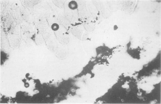
This study was undertaken to determine the sequence of events in the microenvironment of the intestinal tract that culminate in the symptoms of cholera and to attempt to define more clearly the mechanisms involved in antibacterial immunity. The extent to which mucus occurs in the normal intestine of rabbits and the appearance of the intestinal villi in unfixed frozen sections was demonstrated. The villi and intervillous spaces were found to be normally covered by a layer of mucoid material that formed a mucous zone between the intestinal contents and the tips of the villi. The distribution of cholera organisms in normal and immunized animals was demonstrated by the staining of frozen-tissue sections with specific fluorescent antibody. Study of tissue sections from normal animals showed that the onset of fluid accumulation was concomitant with the establishment of large masses of organisms in the intervillous spaces and crypts of the intestine after the successful penetration of this mucous zone. Tissue sections from animals actively or passively immunized against a cell wall antigen of Vibrio cholerae showed clumping of vibrios in the lumen and restricted distribution in the lumen and luminal border of the mucous zone. Antibody was not lytic in vivo.
本研究旨在确定肠道微环境中最终导致霍乱症状的一系列事件,并试图更清楚地界定抗菌免疫所涉及的机制。研究证实了黏液在兔正常肠道中的存在程度以及未固定冷冻切片中肠绒毛的外观。发现绒毛和绒毛间隙通常被一层黏液样物质覆盖,该物质在肠内容物与绒毛顶端之间形成了一个黏液区。通过用特异性荧光抗体对冷冻组织切片进行染色,展示了霍乱弧菌在正常动物和免疫动物中的分布情况。对正常动物组织切片的研究表明,在成功穿透该黏液区后,液体蓄积的开始与大量细菌在肠绒毛间隙和隐窝中的聚集有关。对霍乱弧菌细胞壁抗原进行主动或被动免疫的动物的组织切片显示,弧菌在肠腔内聚集,且在黏液区的肠腔和腔缘分布受限。抗体在体内无溶解作用。