• 文献检索
  • 文档翻译
  • 深度研究
  • 学术资讯
  • Suppr Zotero 插件Zotero 插件
  • 邀请有礼
  • 套餐&价格
  • 历史记录
应用&插件
Suppr Zotero 插件Zotero 插件浏览器插件Mac 客户端Windows 客户端微信小程序
定价
高级版会员购买积分包购买API积分包
服务
文献检索文档翻译深度研究API 文档MCP 服务
关于我们
关于 Suppr公司介绍联系我们用户协议隐私条款
关注我们

Suppr 超能文献

核心技术专利:CN118964589B侵权必究
粤ICP备2023148730 号-1Suppr @ 2026

文献检索

告别复杂PubMed语法,用中文像聊天一样搜索,搜遍4000万医学文献。AI智能推荐,让科研检索更轻松。

立即免费搜索

文件翻译

保留排版,准确专业,支持PDF/Word/PPT等文件格式,支持 12+语言互译。

免费翻译文档

深度研究

AI帮你快速写综述,25分钟生成高质量综述,智能提取关键信息,辅助科研写作。

立即免费体验

霍乱弧菌在实验性霍乱中的黏附和定植:电子显微镜研究

Vibrio cholerae adherence and colonization in experimental cholera: electron microscopic studies.

作者信息

Nelson E T, Clements J D, Finkelstein R A

出版信息

Infect Immun. 1976 Aug;14(2):527-47. doi: 10.1128/iai.14.2.527-547.1976.

DOI:10.1128/iai.14.2.527-547.1976
PMID:971962
原文链接:https://pmc.ncbi.nlm.nih.gov/articles/PMC420916/
Abstract

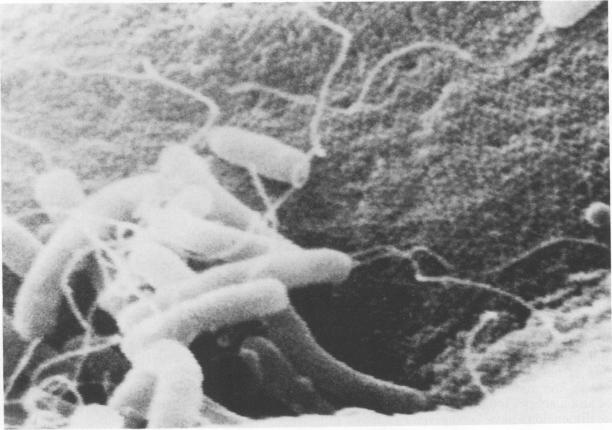
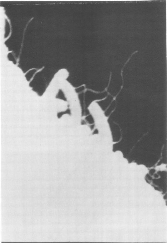
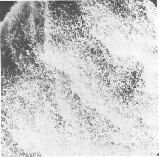
Colonization of the intestinal epithelium by Vibrio cholerae was examined in two model systems, in ligated ileal loops of adult rabbits and in the patent gut of infant rabbits, using both scanning and transmission electron microscopy. Time studies in the adult model showed a lag period of up to 1 h before the attachment of significant numbers of the vibrios. The bacteria appeared initially in small patches on the sides of the villi, predominantly along the transverse furrows. The number of adherent bacteria steadily increased, reaching a maximum between 4 and 7 h, when a dense mat of bacteria several layers thick covered much of the villi. After this time there was a rapid decline in the number of V. cholerae bound. By 12 to 16 h only a few bacteria could be seen on the surface of the villi, which had a rough, patchy appearance at these later times. Globular protrusions, with vibrios attached, may play a role in the clearance of bacteria. Colonization and clearance in the patent intestine of the infant rabbit occurred much as in the adult model. However, the bacteria adhered more uniformly and there was no lag in attachment. In both models the majority of bacteria were aligned horizontally with the epithelial surface, but some were attached in an end-on manner, with their flagella extending into the lumen. The bacteria adhered via their surface coats directly to the tips of the microvilli, except for a few vibrios that were partly embedded into the brush border. Some changes in the microvilli occurred as a consequence of the bacterial attachment.

摘要

利用扫描电子显微镜和透射电子显微镜,在成年兔结扎回肠袢和幼兔开放肠道这两种模型系统中,研究了霍乱弧菌对肠上皮的定殖情况。在成年模型中的时间研究表明,在大量弧菌附着之前有长达1小时的延迟期。细菌最初出现在绒毛侧面的小斑块中,主要沿着横向沟纹分布。附着细菌的数量稳步增加,在4至7小时达到最大值,此时几层厚的密集细菌层覆盖了大部分绒毛。此后,霍乱弧菌的附着数量迅速下降。到12至16小时,在绒毛表面只能看到少数细菌,在后期这些绒毛表面粗糙且呈斑驳状。带有附着弧菌的球状突起可能在细菌清除中起作用。幼兔开放肠道中的定殖和清除情况与成年模型中的情况大致相同。然而,细菌附着更为均匀,且附着没有延迟。在这两种模型中,大多数细菌与上皮表面水平排列,但有些是以端部附着的方式,其鞭毛延伸到肠腔中。除了少数部分嵌入刷状缘的弧菌外,细菌通过其表面被膜直接附着在微绒毛的尖端。由于细菌附着,微绒毛发生了一些变化。

https://cdn.ncbi.nlm.nih.gov/pmc/blobs/027f/420916/c83bded7e4f6/iai00224-0222-a.jpg
https://cdn.ncbi.nlm.nih.gov/pmc/blobs/027f/420916/995e7beaf25d/iai00224-0204-a.jpg
https://cdn.ncbi.nlm.nih.gov/pmc/blobs/027f/420916/d1a17f039272/iai00224-0205-a.jpg
https://cdn.ncbi.nlm.nih.gov/pmc/blobs/027f/420916/dde76393787f/iai00224-0206-a.jpg
https://cdn.ncbi.nlm.nih.gov/pmc/blobs/027f/420916/8270a046d856/iai00224-0207-a.jpg
https://cdn.ncbi.nlm.nih.gov/pmc/blobs/027f/420916/5723cbda1596/iai00224-0208-a.jpg
https://cdn.ncbi.nlm.nih.gov/pmc/blobs/027f/420916/78df03800f0b/iai00224-0209-a.jpg
https://cdn.ncbi.nlm.nih.gov/pmc/blobs/027f/420916/7512e7b09399/iai00224-0210-a.jpg
https://cdn.ncbi.nlm.nih.gov/pmc/blobs/027f/420916/73710bf0e5e6/iai00224-0211-a.jpg
https://cdn.ncbi.nlm.nih.gov/pmc/blobs/027f/420916/4e465cf4cfe4/iai00224-0212-a.jpg
https://cdn.ncbi.nlm.nih.gov/pmc/blobs/027f/420916/13025aaf8b6a/iai00224-0213-a.jpg
https://cdn.ncbi.nlm.nih.gov/pmc/blobs/027f/420916/b3b99592d769/iai00224-0214-a.jpg
https://cdn.ncbi.nlm.nih.gov/pmc/blobs/027f/420916/abcdc4651bf3/iai00224-0215-a.jpg
https://cdn.ncbi.nlm.nih.gov/pmc/blobs/027f/420916/0dd1abcbb3ae/iai00224-0216-a.jpg
https://cdn.ncbi.nlm.nih.gov/pmc/blobs/027f/420916/112d35f50fac/iai00224-0217-a.jpg
https://cdn.ncbi.nlm.nih.gov/pmc/blobs/027f/420916/2709e0f15db7/iai00224-0218-a.jpg
https://cdn.ncbi.nlm.nih.gov/pmc/blobs/027f/420916/7150fbc1e6aa/iai00224-0219-a.jpg
https://cdn.ncbi.nlm.nih.gov/pmc/blobs/027f/420916/f943f21f1216/iai00224-0220-a.jpg
https://cdn.ncbi.nlm.nih.gov/pmc/blobs/027f/420916/b7e95268255a/iai00224-0221-a.jpg
https://cdn.ncbi.nlm.nih.gov/pmc/blobs/027f/420916/c83bded7e4f6/iai00224-0222-a.jpg

相似文献

1
Vibrio cholerae adherence and colonization in experimental cholera: electron microscopic studies.霍乱弧菌在实验性霍乱中的黏附和定植:电子显微镜研究
Infect Immun. 1976 Aug;14(2):527-47. doi: 10.1128/iai.14.2.527-547.1976.
2
In vivo adherence and colonization of Vibrio cholerae strains that differ in hemagglutinating activity and motility.血凝活性和运动性不同的霍乱弧菌菌株在体内的黏附和定殖
Infect Immun. 1987 Sep;55(9):2093-102. doi: 10.1128/iai.55.9.2093-2102.1987.
3
Interaction of vibrio cholerae El Tor and gut mucosa in ligated rabbit ileal loop experiment.霍乱弧菌埃尔托生物型与兔回肠结扎肠袢实验中肠道黏膜的相互作用
Med Biol. 1977 Jun;55(3):130-40.
4
Localization of Vibrio cholerae O1 in the intestinal tissue.霍乱弧菌O1在肠道组织中的定位
Asian Pac J Allergy Immunol. 1993 Dec;11(2):155-65.
5
Suppression of growth of Vibrio cholerae in the intestine of immunized mice.免疫小鼠肠道中霍乱弧菌生长的抑制
Med Microbiol Immunol. 1976 Jun 1;162(2):153-8. doi: 10.1007/BF02121325.
6
Electron microscopic study of Vibrio cholerae O1 adherence to the mucus coat and villus surface in the human small intestine.霍乱弧菌O1对人小肠黏液层和绒毛表面黏附的电子显微镜研究。
Infect Immun. 1988 Oct;56(10):2753-9. doi: 10.1128/iai.56.10.2753-2759.1988.
7
Successful colonization and immunization of adult rabbits by oral inoculation with Vibrio cholerae O1.通过口服接种霍乱弧菌O1对成年兔进行成功定殖和免疫。
Infect Immun. 1983 Aug;41(2):735-41. doi: 10.1128/iai.41.2.735-741.1983.
8
[Quantitative evaluation of Vibrio cholerae colonization and lymphocyte count in small intestine villi of rabbits immunized with vibrio and cholera toxin].
Zh Mikrobiol Epidemiol Immunobiol. 1982 Feb(2):63-70.
9
RpoS controls the Vibrio cholerae mucosal escape response.RpoS控制霍乱弧菌的黏膜逃逸反应。
PLoS Pathog. 2006 Oct;2(10):e109. doi: 10.1371/journal.ppat.0020109.
10
Vibrio cholerae O1 adherence to villi and lymphoid follicle epithelium: in vitro model using formalin-treated human small intestine and correlation between adherence and cell-associated hemagglutinin levels.霍乱弧菌O1对绒毛和淋巴滤泡上皮的黏附:使用经福尔马林处理的人小肠的体外模型以及黏附与细胞相关血凝素水平之间的相关性
Infect Immun. 1988 Dec;56(12):3241-50. doi: 10.1128/iai.56.12.3241-3250.1988.

引用本文的文献

1
The Interface of and the Gut Microbiome.和肠道微生物组的界面。
Gut Microbes. 2021 Jan-Dec;13(1):1937015. doi: 10.1080/19490976.2021.1937015.
2
Host-Microbe-Pathogen Interactions: A Review of Pathogenesis in .宿主-微生物-病原体相互作用:对 发病机制的综述。
Front Immunol. 2020 Jan 24;10:3128. doi: 10.3389/fimmu.2019.03128. eCollection 2019.
3
Anti-biofilm Properties of the Fecal Probiotic Lactobacilli Against spp.粪便益生菌乳杆菌对 spp. 的抗生物膜特性

本文引用的文献

1
The fate of bacteria in the small intestine.小肠中细菌的命运。
J Pathol Bacteriol. 1960 Jan;79:131-40. doi: 10.1002/path.1700790116.
2
Factors of virulence of Vibrio cholerae.霍乱弧菌的毒力因子。
Ann N Y Acad Sci. 1960 Nov 21;88:1203-12. doi: 10.1111/j.1749-6632.1960.tb20111.x.
3
Minimum nutritional requirements for mucinase synthesis by Vibrio cholerae.
Tex Rep Biol Med. 1960;18:612-9.
Front Cell Infect Microbiol. 2018 Apr 24;8:120. doi: 10.3389/fcimb.2018.00120. eCollection 2018.
4
Vibrio cholerae Biofilms and Cholera Pathogenesis.霍乱弧菌生物膜与霍乱发病机制
PLoS Negl Trop Dis. 2016 Feb 4;10(2):e0004330. doi: 10.1371/journal.pntd.0004330. eCollection 2016 Feb.
5
Bacterial community associated with the intestinal tract of Chinese mitten crab (Eriocheir sinensis) farmed in Lake Tai, China.与中国太湖养殖的中华绒螯蟹(Eriocheir sinensis)肠道相关的细菌群落。
PLoS One. 2015 Apr 13;10(4):e0123990. doi: 10.1371/journal.pone.0123990. eCollection 2015.
6
Manipulation of intestinal epithelial cell function by the cell contact-dependent type III secretion systems of Vibrio parahaemolyticus.通过副溶血性弧菌的细胞接触依赖性 III 型分泌系统对肠上皮细胞功能的操纵。
Front Cell Infect Microbiol. 2014 Jan 10;3:114. doi: 10.3389/fcimb.2013.00114. eCollection 2014.
7
Tn-Seq analysis of Vibrio cholerae intestinal colonization reveals a role for T6SS-mediated antibacterial activity in the host.Tn-Seq 分析霍乱弧菌肠道定植揭示 T6SS 介导的抗菌活性在宿主中的作用。
Cell Host Microbe. 2013 Dec 11;14(6):652-63. doi: 10.1016/j.chom.2013.11.001.
8
Stringent response regulation of biofilm formation in Vibrio cholerae.霍乱弧菌生物膜形成的严格响应调控。
J Bacteriol. 2012 Jun;194(11):2962-72. doi: 10.1128/JB.00014-12. Epub 2012 Mar 30.
9
Inflammation and disintegration of intestinal villi in an experimental model for Vibrio parahaemolyticus-induced diarrhea.实验性创伤弧菌性腹泻模型中肠绒毛的炎症和崩解。
PLoS Pathog. 2012;8(3):e1002593. doi: 10.1371/journal.ppat.1002593. Epub 2012 Mar 15.
10
Protection and attachment of Vibrio cholerae mediated by the toxin-coregulated pilus in the infant mouse model.毒素调节菌毛介导的婴儿鼠模型中霍乱弧菌的保护和黏附。
J Bacteriol. 2011 Oct;193(19):5260-70. doi: 10.1128/JB.00378-11. Epub 2011 Jul 29.
4
An evaluation of intestinal fluids in the pathogenesis of cholera.霍乱发病机制中肠液的评估。
J Infect Dis. 1961 Jul-Aug;109:35-42. doi: 10.1093/infdis/109.1.35.
5
An experimental study of the mechanism of action of Vibriod cholerae on the intestinal mucous membrane.霍乱弧菌对肠黏膜作用机制的实验研究
J Pathol Bacteriol. 1953 Oct;66(2):559-62. doi: 10.1002/path.1700660228.
6
Experimental cholera. Morphologic evidence of cytotoxicity.
Arch Pathol. 1966 Jul;82(1):54-9.
7
Pathogenesis of experimental cholera: identification of choleragen (procholeragen A) by disc immunoelectrophoresis and its differentiation from cholera mucinase.实验性霍乱的发病机制:通过圆盘免疫电泳鉴定霍乱毒素原(前霍乱毒素A)及其与霍乱粘蛋白酶的鉴别
J Immunol. 1966 Jul;97(1):25-33.
8
Small bowel morphology in experimental canine cholera. A light and electron microscopic study.实验性犬霍乱的小肠形态学。光镜与电镜研究。
Lab Invest. 1970 Feb;22(2):112-20.
9
Electron microscopy of the small intestine: a review.
Gastroenterology. 1965 Nov;49(5):574-603.
10
Goblet cell mucus in the small intestine. Findings after net fluid production due to cholera toxin and hypertonic solutions.小肠中的杯状细胞黏液。霍乱毒素和高渗溶液导致净液体产生后的发现。
Johns Hopkins Med J. 1972 Jul;131(1):1-10.