Evans Mark F, Aliesky Holly A, Cooper Kumarasen
Department of Pathology, University of Vermont, Burlington, VT 05405, USA.
BMC Clin Pathol. 2003 Jun 11;3(1):2. doi: 10.1186/1472-6890-3-2.
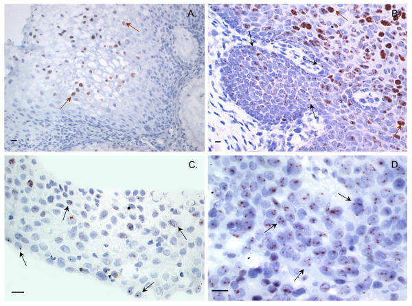
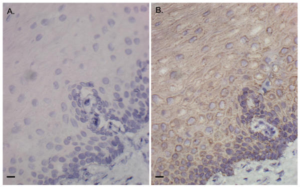
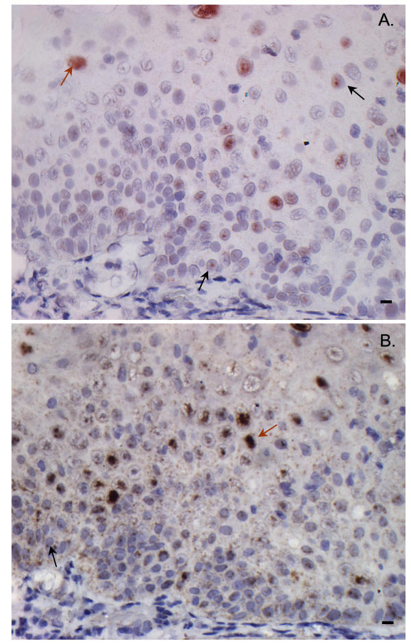
Over the past five years in situ hybridization techniques employing tyramide amplification reagents have been developed and promise the potential detection of low/single-copy nucleic acid sequences. However the increased sensitivity that tyramide amplification brings about may also lead to problems of background staining that confound data interpretation. METHODS: In this study those factors enabling background-free biotinyl-tyramide based in situ hybridization assay of formalin-fixed paraffin-embedded tissues have been examined. SiHa, HeLa and CaSki cell lines known to contain HPV integrated into the cell genome, and archival cervical pre-invasive lesions and carcinomas have been successfully assessed using biotinylated HPV and centromeric probes. RESULTS: The single most important factor both for sensitivity and clean background was a tissue unmasking regimen that included treatment with 10 mM sodium citrate pH 6.0 at 95 degrees C followed by digestion with pepsin/0.2 M HCl. Concentrations both of probe and primary streptavidin-peroxidase conjugate and pH of hybridization mix and stringency washes were also critical for sensitivity. Certain probes were more associated with background staining than others. This problem was not related to probe purity or size. In these instances composition of hybridization mix solution was especially critical to avoid background. 3-amino-9-ethylcarbazole was preferred over 3,3'-diaminobenzidene as a chromogen because background was cleaner and the 1-2 copies of HPV16 integrated in SiHa cells were readily demonstrable. HPV detection on metaphase spreads prepared from SiHa cells was only successful when a fluorescent detection method was combined with tyramide reagent. 'Punctate' and 'diffuse' signal patterns were identified amongst tissues consistent with the former representing integration and 'diffuse' representing episomal HPV. Only punctate signals were detected amongst the cell lines and were common amongst high-grade pre-invasive lesions and carcinomas. However it remains to be determined why single/low-copy episomal HPV in basal/parabasal cells of low-grade lesions is not also detectable using tyramide-based techniques and whether every punctate signal represents integration. CONCLUSIONS: A tyramide-based in situ hybridization methodology has been established that enables sensitive, background-free assay of clinical specimens. As punctate signals characterize HPV in high-grade cervical lesions the method may have potential for clinical applications.
在过去五年中,已开发出采用酪胺扩增试剂的原位杂交技术,有望检测低拷贝/单拷贝核酸序列。然而,酪胺扩增带来的灵敏度提高也可能导致背景染色问题,从而混淆数据解读。方法:在本研究中,对那些能够实现福尔马林固定石蜡包埋组织基于生物素化酪胺的无背景原位杂交检测的因素进行了研究。已知含有整合到细胞基因组中的人乳头瘤病毒(HPV)的SiHa、HeLa和CaSki细胞系,以及存档的宫颈癌前病变和癌组织,已使用生物素化HPV探针和着丝粒探针成功进行了评估。结果:对于灵敏度和清晰背景而言,最重要的单一因素是组织预处理方案,包括在95℃下用10 mM pH 6.0柠檬酸钠处理,然后用胃蛋白酶/0.2 M盐酸消化。探针浓度、一级链霉亲和素-过氧化物酶缀合物浓度、杂交混合液的pH值和严谨性洗涤对灵敏度也至关重要。某些探针比其他探针更容易出现背景染色。这个问题与探针纯度或大小无关。在这些情况下,杂交混合液的组成对于避免背景尤其关键。作为显色剂,3-氨基-9-乙基咔唑比3,3'-二氨基联苯胺更受青睐,因为背景更干净,并且在SiHa细胞中整合的1至2个HPV16拷贝很容易被检测到。只有当荧光检测方法与酪胺试剂结合时,从SiHa细胞制备的中期染色体铺片上的HPV检测才成功。在组织中识别出了“点状”和“弥漫性”信号模式,前者代表整合,“弥漫性”代表游离型HPV。在细胞系中仅检测到点状信号,在高级别癌前病变和癌组织中很常见。然而,仍有待确定为什么使用基于酪胺的技术无法检测低级别病变的基底/副基底细胞中的单拷贝/低拷贝游离型HPV,以及每个点状信号是否都代表整合。结论:已建立了一种基于酪胺的原位杂交方法,可对临床标本进行灵敏、无背景的检测。由于点状信号是高级别宫颈病变中HPV的特征,该方法可能具有临床应用潜力。